Fig. 2:
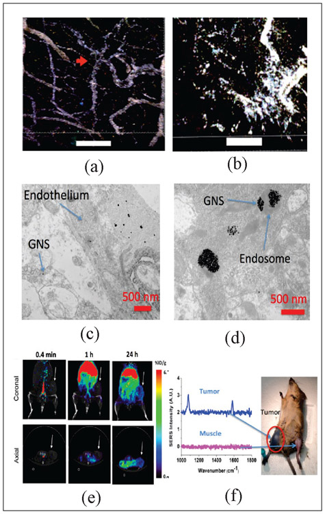
(a-b) Two-photon photoluminescence image of blood-brain barrier interface of glioblastoma model on mice treated with GNS and PTT (scale bar: 100 μm). GNS (white) initially only inside blood vessels, but after some time they are found extravasating into the surrounding parenchyma (Adapted from Ref. 38). (a) Image just before treatment. The red arrow denotes vascular tortuosity. (b) Image 48 hours after laser treatment. (c) Electron microscopy also shows that GNS nanoparticles penetrate through brain tumor vasculature and (d) can get inside the brain cancer cell (Adapted from Ref. 39). (e) PET/CT of GNS 64Cu 0.4 min, 1h and 24h after IV injection. The GNS accumulate in tumor over time (Adapted from Ref. 6) (f) SERS spectra of PMBA labeled GNS in tumor flank compared to SERS at nearby muscle tissue (Adapted from Ref. 16)

