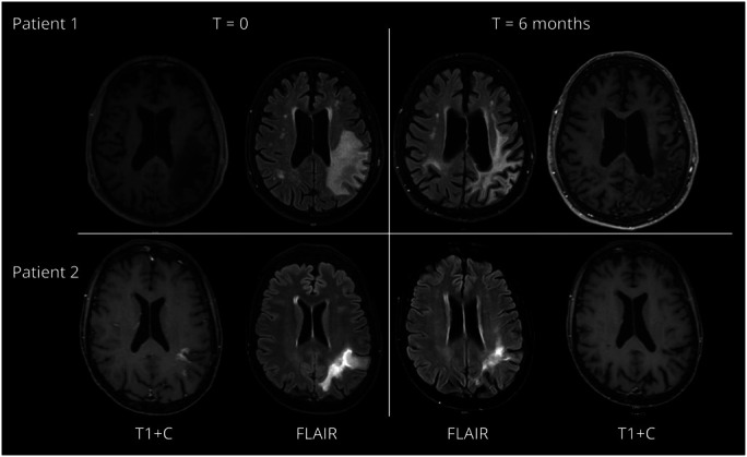
Figure. Axial MRI FLAIR and Contrast-Enhanced T1 Images (Radiologic Convention, Left = Right) of the 2 Patients at Baseline (T = 0) and after 6 months (T+6 M).

In patient 1 (upper row), at baseline, multiple hyperintense lesions on the fluid-attenuated inverse recovery (FLAIR) sequence were visible. The largest was located in the left parietal cortex. The lesions included the U fibers. After IV gadolinium, no contrast enhancement was visible. At ±6 months, the size of the lesions had decreased and atrophy was visible in the affected areas. In patient 2 (lower row), at baseline, multiple hyperintense lesions on the FLAIR sequence were visible. The largest was located in the left parietal cortex. The lesions included the U fibers. After IV gadolinium, ring enhancement was visible. At ±6 months, the lesions were less outspoken and no contrast enhancement was visible anymore.
