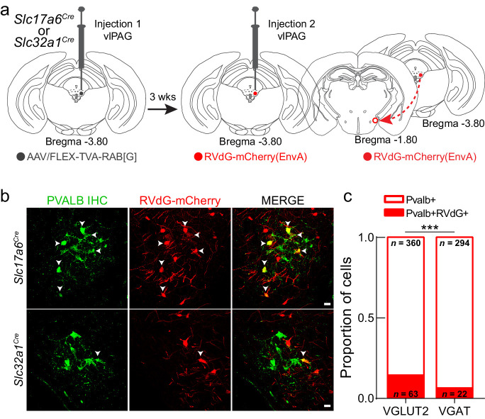
Figure 5. LHPV neurons preferentially target glutamatergic neurons in the ventrolateral periaqueductal gray area (vlPAG).
(a) Schematic for modified rabies viral tracing strategy. (b) Images from Slc17a6Cre (top row) and Slc32a1Cre (bottom row) brain slices showing the overlap of RVdG-mCherry(EnvA) with LHPV neurons. Scale bars: 20 μm. (c) Proportion of LHPV neurons that express or do not express RVdG-mCherry(EnvA) in Slc17a6Cre or Slc32a1Cre mice. LHPV neurons were connected to a greater proportion of vlPAGVGLUT2 neurons than vlPAGVGAT neurons (chi-square = 11.18, p=0.0008).

