. 2020 Apr 20;11(29):7641–7653. doi: 10.1039/d0sc01949f

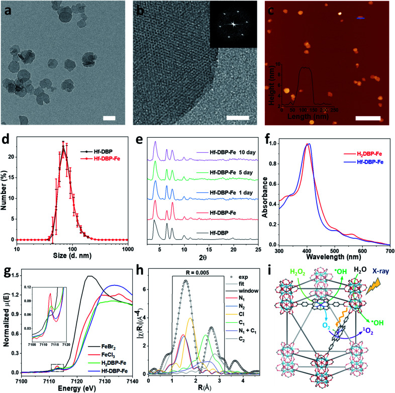
Fig. 2. Characterization of Hf-DBP-Fe (a) TEM image and (b) HRTEM image with the FFT pattern (inset) of Hf-DBP-Fe. (c) AFM topography and its height profile (inset) along the blue line of Hf-DBP-Fe. (d) Number-averaged diameters of Hf-DBP-Fe and Hf-DBP in ethanol. (e) PXRD pattern and stability test of Hf-DBP-Fe. (f) UV-Vis spectra of Hf-DBP-Fe in comparison to H2DBP-Fe. (g) XANES analysis of Hf-DBP-Fe and H2DBP-Fe showing the pre-edge feature of FeIII in both Hf-DBP-Fe and H2DBP-Fe. (h) EXAFS fitting of Hf-DBP-Fe to five-coordinate porphyrin-FeIIICl as shown in Fig. S1. (i) Schematic showing Hf-DBP-Fe-mediated CDT and RT-RDT processes. The DBP-Fe centers catalyze the generation of ˙OH (CDT) and O2 from H2O2. Upon X-ray irradiation, Hf-DBP-Fe enhances radiosensitization to produce additional ˙OH (RT) and convert the generated O2 into 1O2 (RDT). Scale bar = 100 nm in a, 20 nm in b, and 500 nm in c, respectively.

Fig. 2