. 2020 Apr 7;11(17):4397–4402. doi: 10.1039/d0sc00844c

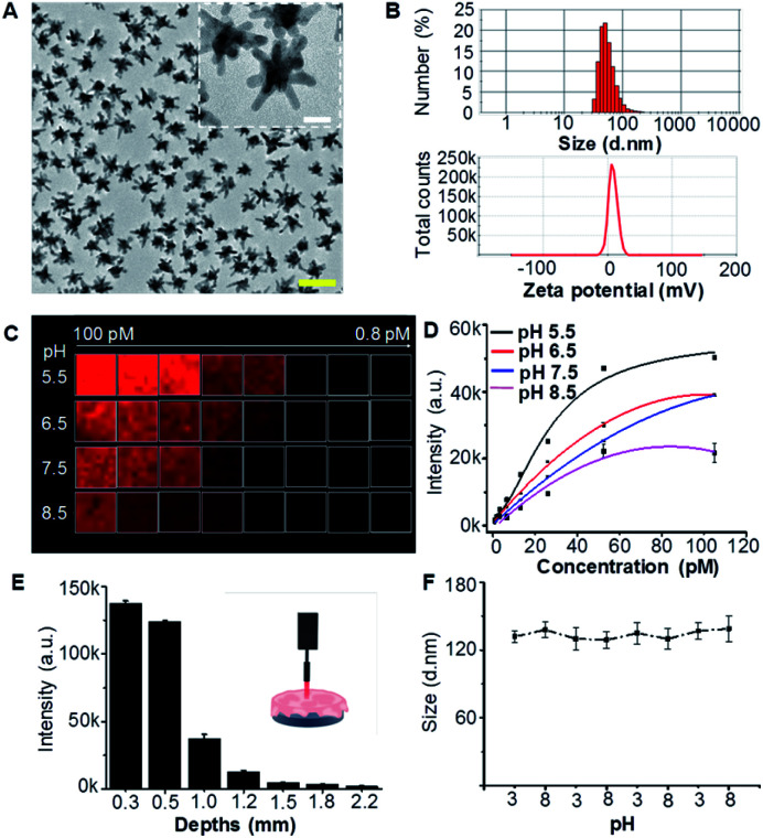
Fig. 2. Characterization of AuS-IR7p. (A) TEM image of AuS-IR7p. Scale bar = 100 nm. Inset: amplified gold nanostars. Scale bar = 20 nm. (B) Hydrodynamic diameter distribution and zeta potentials of AuS-IR7p in PBS solution (pH 7.4). (C) Raman microscopy image of AuS-IR7p as functions of concentrations and pH values by quantifying its characteristic peak at 510 cm−1. (D) Concentration dependent Raman signal intensities of AuS-IR7p in buffered solutions with different pH values. (E) Raman signal intensities of AuS-IR7p as a function of the thickness of covered mouse brain tissue (200 mW laser power density, 1.0 s acquirement time, and 10× objective). Data are presented as mean ± S.D. (n = 3). (F) AuS-IR7p shows reversible pH responsive ability. The initial concentration was 100 pM. Laser power: 80 mW. Acquirement time: 1.0 s.

Fig. 2