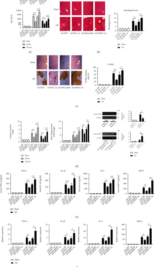
Figure 2.

PGC-1α protects the liver against I/R injury. (a) Serum levels of aminotransferases (ALT and AST) were detected in the mice subjected to Ad-GFP, Ad-PGC-1α, Ad-shScramble, and Ad-shPGC-1α at 6 h and 24 h after liver I/R (n = 6). (b) Representative images (200x magnification) of H&E-stained liver sections (6 h after I/R) were taken, and histopathological scoring of hepatic injury was performed. (c) Representative images (200x magnification) of liver sections (6 h after I/R) stained by TUNEL were taken, and TUNEL-positive cells were counted as described in Materials and Methods. (d) Caspase-3 activity, DNA fragmentation, and cleaved PARP expression in the mouse livers were assessed by ELISA and western blot (n = 3-6). (e) Systemic TNF-α, IL-1β, IL-6, and MIP-2 levels at 6 h after liver I/R were measured by ELISA (n = 6). (f) The relative mRNA expression levels of TNF-α, IL-1β, IL-6, and MIP-2 in the mouse liver tissues at 6 h after I/R were determined by quantitative RT-PCR (n = 3). ∗P < 0.05, ∗∗P < 0.01, and ∗∗∗P < 0.001.
