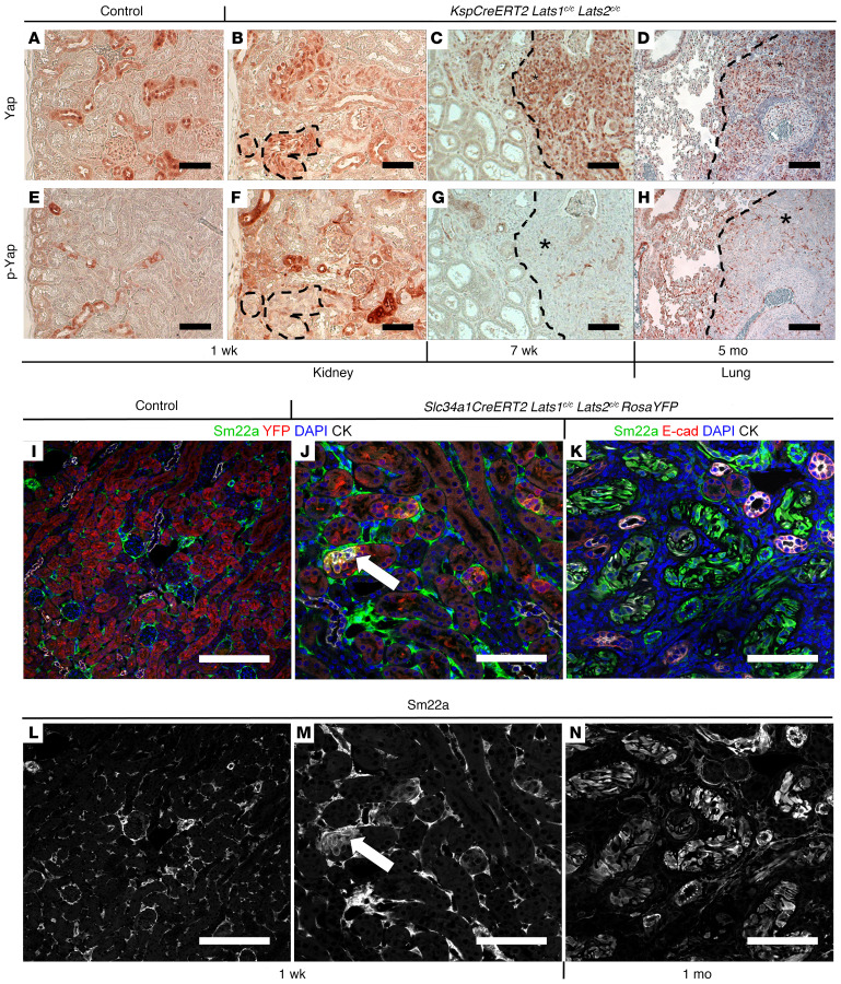
Figure 3. Molecular analysis of Lats mutants.
(A–H) Immunohistochemical staining against Yap (A–D) or p-Yap (E–H) on sections of control kidney (A and E; n = 3), KspCreERT2 Lats1c/c Lats2c/c kidney 1 week (B and F; n = 3) or 7 weeks after tamoxifen (C and G; n = 3), and KspCreERT2 Lats1c/c Lats2c/c lung 5 months after tamoxifen (D and H; n = 3). (I–N) Immunofluorescence staining on control (I and L) or Slc34a1CreERT2 Lats1/2c/c RosaYFP kidneys (J, K, M, and N) 1 week (J and M; n = 3 control, n = 5 mutant) or 1 month (K and N; n = 4) after tamoxifen. (I and J) Green, Sm22a; red, YFP; blue, DAPI; white, cytokeratin. (K) Green, Sm22a; red, E-cadherin; blue, DAPI; white, cytokeratin. L–N are single-channel images of Sm22a from I–K, respectively. All scale bars: 100 μm.

