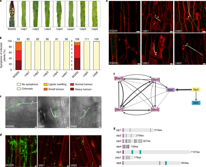
Fig. 1. Seven U. maydis proteins essential for virulence form a protein complex.
a,b, Seven-day-old maize seedlings were infected with SG200 and the indicated deletion strains. At 12 d.p.i., representative leaves were photographed (a) and disease symptoms were scored (b). The vertical dashed line separates two independent sets of experiments. Data represent the mean of n = 3 biologically independent experiments. Total numbers of infected plants are indicated above the respective columns. c, Maize epidermal cells 1 d.p.i. with SG200AN1, SG200AM1∆stp1 and SG200AN1∆stp2 all expressing cytosolic GFP upon plant penetration. The GFP (green) and bright-field (grey) channels are merged. Hyphae on the plant surface are traced in white; untraced hyphae are intracellular. Arrowheads indicate appressoria. Shown are maximum projections of confocal z-stacks. Scale bars, 5 μm. d, Maize leaves 3 d.p.i. with SG200 and the indicated deletion strains stained with WGA-AF488 (fungal cell wall, green) and propidium iodide (plant cell wall, red). Shown are AF488 (green) and propidium iodide (red) channel overlays of confocal z-stack maximum projections. Scale bars, 25 μm. e, Maize epidermal cells 1 d.p.i. (upper panel) and 2 d.p.i. (lower panel) with SG200AN1, SG200AM1∆stp1 and SG200AN1∆stp2 all expressing cytosolic GFP upon plant penetration. Plasma membranes were stained with FM4-46 (red). Shown are GFP (green) and FM4-46 (red) channel overlays of confocal z-stack maximum projections. Hyphae on the plant surface are traced in white; intracellular hyphae are untraced. Arrowheads indicate appressoria. Scale bars, 10 μm. f, Effector protein complex interaction network resulting from co-IP/MS experiments using Stp1, Stp2, Stp3, Stp4, Pep1 and Stp6 as bait proteins across several replicated experiments. Line widths illustrate the average number of spectral counts across experiments. g, Domain arrangement of the Stp proteins and calculated molecular weight without signal peptide. Violet, signal peptide; black vertical lines, cysteine residues; green, transmembrane domains predicted based on sequence conservation to membrane domains containing orthologues from other smut fungi (Supplementary Figs. 1–7).

