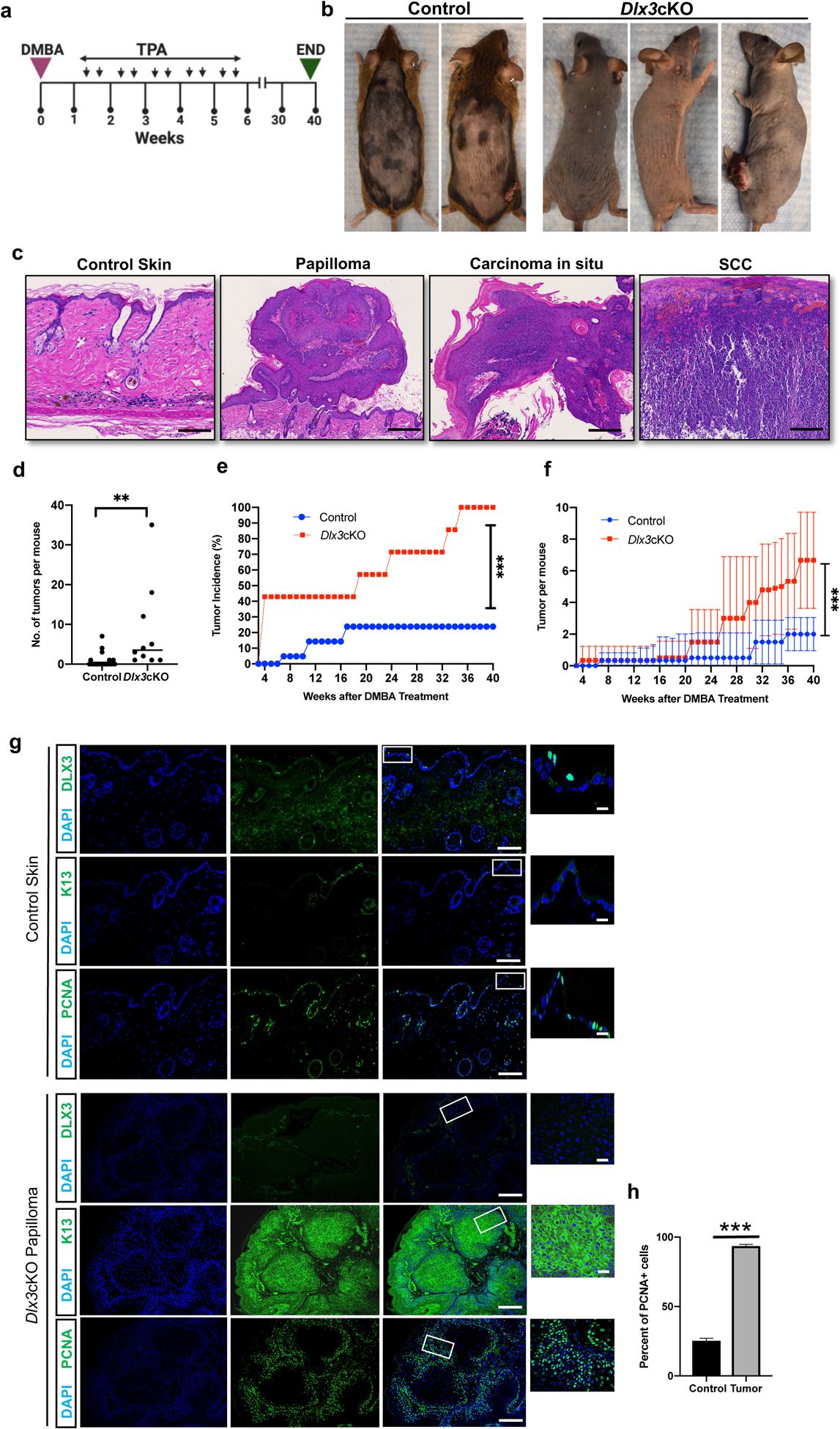
Figure 2. Dlx3 deficiency enhances the development of DMBA/TPA-induced skin tumors.

(a) Experimental design for a chemically induced skin tumorigenesis study. After DMBA treatment, mice were applied with TPA twice a week for 5 weeks. Tumors larger than 1 mm in diameter were counted weekly. At week 40, tumors were collected for analysis. Dlx3cKO; n=11: Control; n=23 (b) Dorsal view of control and Dlx3cKO mice at week 40. (c) H&E staining of wild type skin, papilloma, carcinoma in situ and SCC (Scale bar, 100μm). (d) Number of tumors per mouse. (e) Tumor incidence; percentage (%) of mice that developed any tumors. (f) Average number of tumors per mouse in the total number of mice tested. (g) Representative immunohistochemistry of DLX3, K13 and PCNA (Scale bars, 100μm and 10μm). As shown DLX3 expression is absent in the tumors. Higher PCNA expression and elevated K13 expression in cytoplasm was detected in tumors from Dlx3cKO mice. (h) Detection of proliferating cell nuclear antigen (PCNA)-positive cells in tumor tissue with the control skin. Data are presented as mean ± standard error of the mean. (*** P-value <0.001). n=number of mice.
