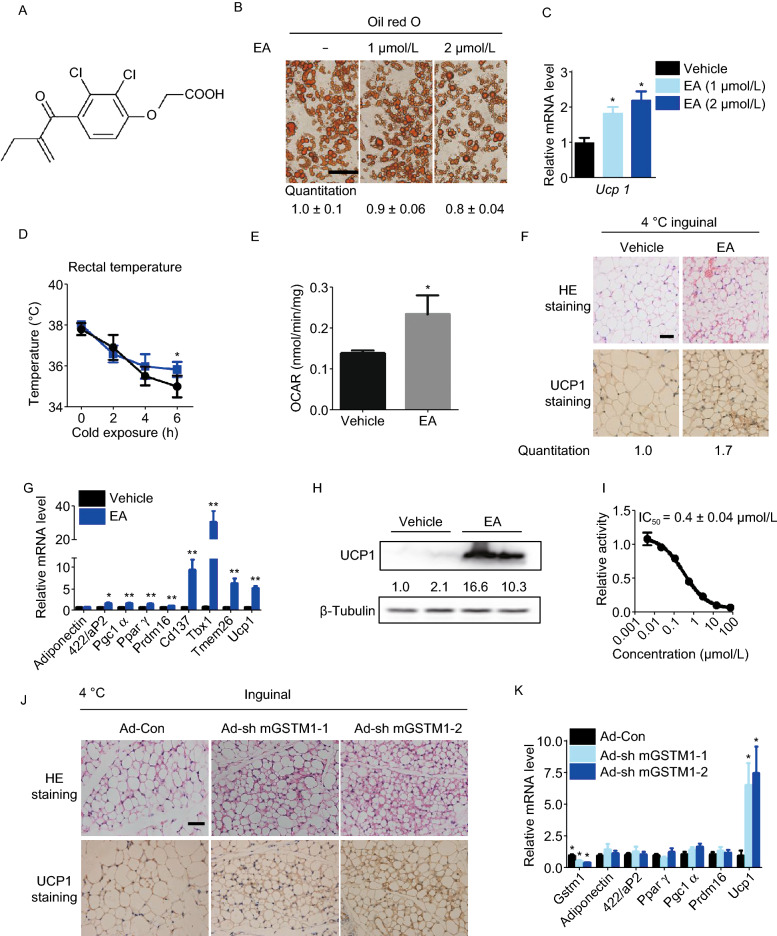
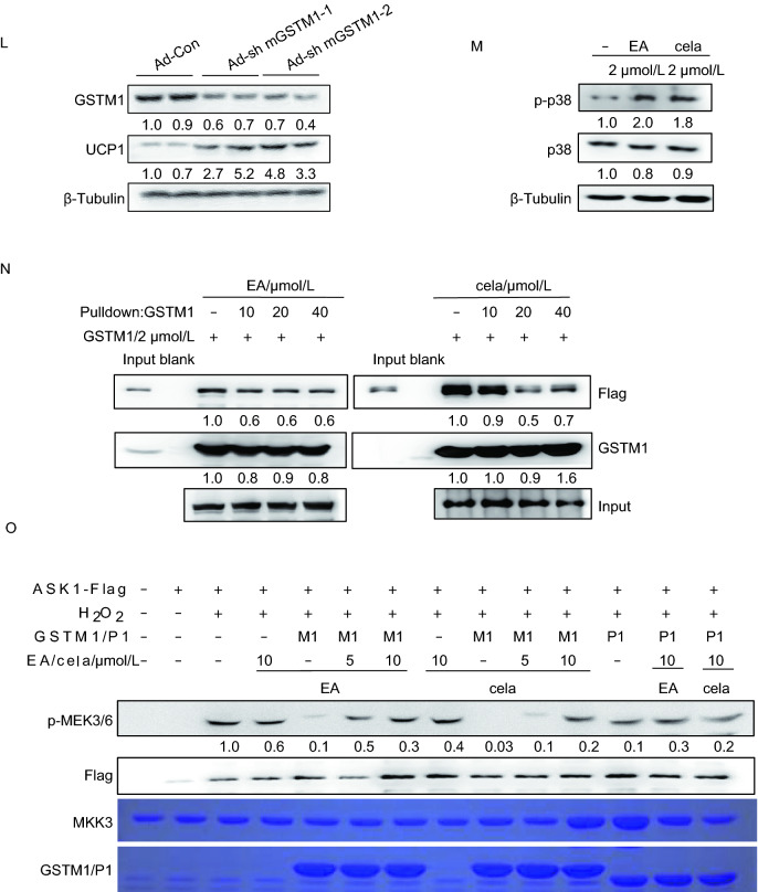
Figure 1.
Ethacrynic acid and celastrol promote browning through GSTM1-ASK1-p38 MAPK pathway. (A) Chemical structure of ethacrynic acid. (B) Oil red O staining at microscopic levels in C3H10T1/2 cells with or without EA treatment during adipogenesis. Average size of lipid droplets in cells were quantified and showed below the figure. Scale bar 50 μm. (C) Transcriptional levels of Ucp1 in C3H10T1/2 with or without EA treatment during adipogenesis. (D) Rectal body temperature of control and EA-treated mice maintained on HFD during cold exposure (4 °C); n = 6 per group. (E) Oxygen consumption rates of inguinal adipose tissues in mice with or without EA treatment for 1 week; n = 5 per group. (F) Representative HE staining and IHC staining of UCP1 in the inguinal adipose tissue of HFD-fed mice after treatment with EA for 1 week and then exposed to 4 °C for 8 h. The UCP1 positive area were quantified and showed below. (G) Transcriptional levels of browning-relevant genes in iWAT of HFD-fed mice after EA administration and cold exposure. (H) UCP1 expression level in iWAT of HFD-fed mice after treatment with EA for 1 week exposed to 4 °C for 8 h. (I) Effect of EA on GSTM1 catalytic activity. (J) Representative IHC staining of UCP1 and HE staining with or without GSTM1 knockdown in iWAT of mice with cold exposure at 4 °C for 8 h, n = 5. Scale bar, 50 µm. The UCP1 positive area were quantified and showed below. (K) mRNA expression of related genes with or without GSTM1 knockdown in iWAT of mice with cold exposure at 4 °C for 8 h, n = 5. (L) Western blot analysis of UCP1 levels with or without GSTM1 knockdown in iWAT of mice with cold exposure at 4 °C for 8 h, n = 5. (M) Protein levels of p-p38, and p38 in mature C3H10T1/2 with cela or EA treatment for 3 h. (N) In vitro effects of EA/cela on the interaction between GSTM1 and ASK1, performed in duplicate. (O) 293T cells were transfected with pcDNA3.1-ASK1-FLAG and treated with H2O2 (2 mmol/L, 20 min) after 36 h of transfection. The cell lysates were subsequently subjected to immol/Lunoprecipitation with anti-FLAG antibody. The immunopellets were then evaluated for ASK1 activity with immunocomplex kinase assay in the presence of GSTM1 (2 mmol/L) and indicated concentrations of cela and EA. Unless specifically stated, values are presented as mean ± s.e.m from three independent experiments and *P < 0.05, **P < 0.01, ***P < 0.001 compared to control groups, as determined by student’s t test


