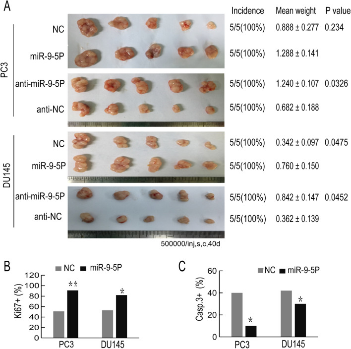
Figure 6.
Overexpression of miR-9-5p promotes xenograft tumor growth in vivo. (A) Overexpression or decreased expression of miR-9-5p in PC3 or DU145 cells was generated, after which the transfected cells were subcutaneously injected into BALB/C nude mice. Tumors were harvested and the tumor images is shown on the left of the picture. The incidence (tumors/injections) and weight of the tumors (mean ± S.D.) and the corresponding P values are shown on the right of the picture. (B, C) Representative immunohistochemical (IHC) staining pictures are shown, in which the Ki-67- or Caspase-3-positive cells from the harvested tumor tissues were stained (magnification, × 200). The percentage of Ki67-positive or Caspase-3-positive cells was calculated. All results are represented as the mean ± SEM from three independent assays. *P < 0.05, **P < 0.01.

