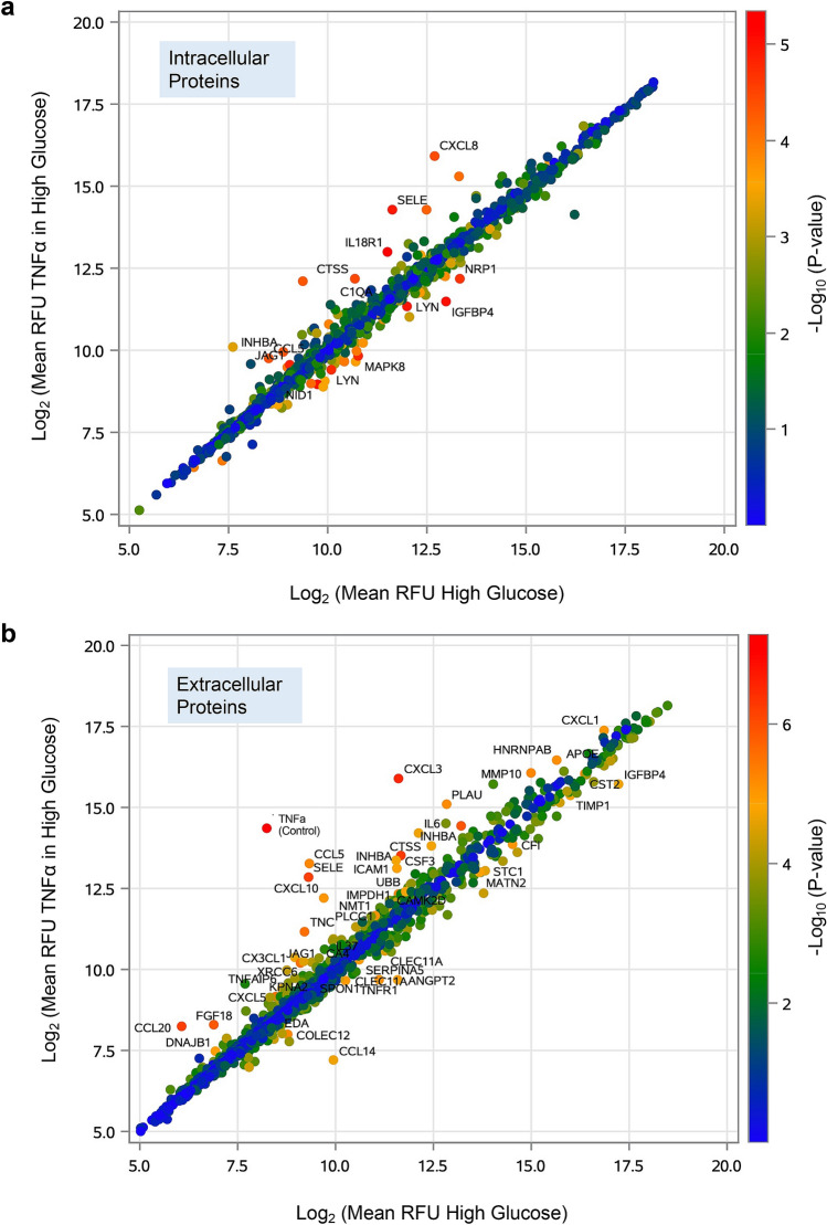
Figure 2.
Protein expression profiles in HUVECs exposed to TNFα in hyperglycemia condition vs. hyperglycemia alone. Scatterplots comparing (a) intracellular and (b) extracellular protein expression profiles in HUVECs exposed to TNFα (10 ng/mL) in high glucose vs. high glucose (4.5 g/L) alone. The values plotted are the mean RFU values (log2 scaled for 3 replicates) for the TNF-α in high glucose (y axis) and the high glucose (x axis) groups. The color of each point indicates the P-values intensity (-log10 scaled) from not significant (blue) to highly significant (red). Intracellular (n = 14) and extracellular (n = 48) proteins with Bonferroni’s corrected α = 3.8 × 10⁻⁵ (0.05/1305) are indicated on the plots.

