Figure 1.
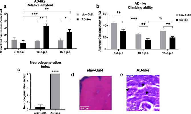
AD-like flies show a degenerative phenotype. (a) Amyloid quantification through Thioflavin aggregation assay. AD-like flies presented a higher amyloid content when compared to control flies (elav-Gal4) at 10- and 15-days post eclosion (d.p.e.) (n = 20 in each genotype). (b) Climbing ability. AD-like flies presented a lower climbing ability in the RING assay than the control genotype at 5–8, 10–13, and 15–18 days post eclosion. AD-like flies present a climbing ability decrease when comparing flies at 5–8 and 10–13 d.p.e. (n ≥ 180 in each genotype). (c) Neurodegeneration index of elav-Gal4 (control) and AD-like flies based on histopathology analysis, according to vacuolar lesions. 0 indicates no lesions, and 5 indicates neurodegenerative phenotype (n = 10, at 10 days after treatment). Data are shown as the mean ± S.E.M. The statistical significance is indicated as ** for P < 0.01 and *** for P < 0.001 (two-tailed ANOVA). Illustrative images of (d) elav-Gal4 and (e) AD-like flies’ histopathology. Arrows indicate vacuolar lesions.

