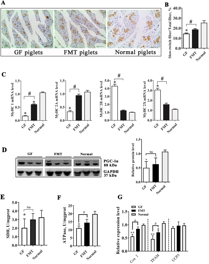
Figure 7.
Change in the muscle fiber type in GF piglets. (A) Immunohistochemical analysis of muscle samples. The brown spots indicate slow-twitch muscle fibers. (B) Proportion of slow-twitch muscle fibers in the total muscle fibers. The full-length blots are presented in Supplementary Fig. 2. (C) mRNA levels of different MyHC genes in the muscle tissues of piglets were detected by qRT-PCR. (D) Protein level of PGC-1α and relative quantitative analysis of the protein level. (E) Enzyme activity of succinate dehydrogenase (SDH) in muscle. (F) Enzyme activity of Na+K+-ATPase in muscle. (G) Changes in the expression of the cytochrome c oxidase I (Cox I), transcription factor A mitochondrial (TFAM), and uncoupling protein 3 (UCP3) genes in muscle tissue. N = 3, the data are presented as the means ± s.e.m. *P < 0.05, **P < 0.01 compared to the normal piglets (control). #P < 0.05 between the GF piglets and FMT piglets. NS not significant, GF piglets germ-free piglets, FMT piglets GF piglets with fecal microbiota transplantation.

