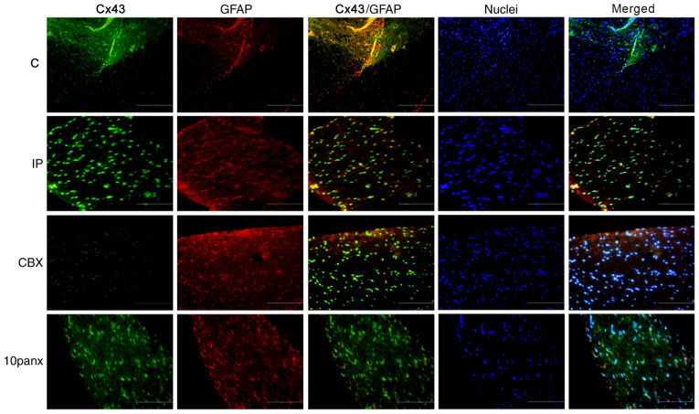
Figure 4.
Co-localization of Cx43 and GFAP in IP model rats. Immunofluorescence assay was used to evaluate the co-localization of Cx43 (green) and GFAP (red). Magnification, ×200; scale bar, 10 µm. C, control group; IP, incision pain; CBX, carbenoxolone; 10panx, pannexin-1 mimetic inhibitory peptide; Cx43, connexin 43; GFAP, glial fibrillary acidic protein.

