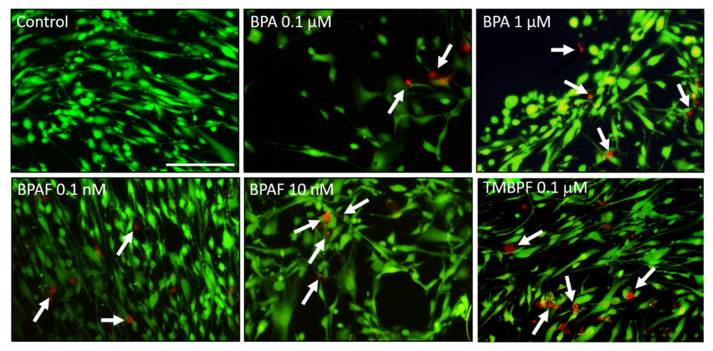
Figure 2.
Cytotoxicity assay of BPA and BPA alternatives. Representative fluorescence images of hASCs treated with BPA at 0.1 µM and 1 µM, BPAF at 0.1 nM and 10 nM, or with TMBPF at 0.1 µM, after 24 h of exposure. Green indicates live cells and red indicates dead cells. Some cell death (see arrows) was found at each dose, especially with 0.1 μM TMBPF, but in general cells remained viable after 24 h of exposure to low-dose BPA and BPAF (200× magnification; scale bar = 150 μM; n = 3–4 slides/treatment; 3 trials).

