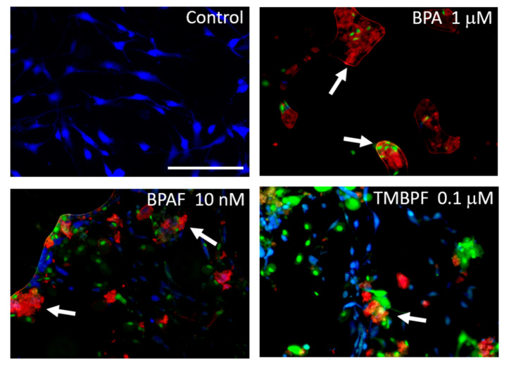
Figure 6.
BPA and alternatives increase apoptosis. Representative fluorescent images of control, BPA-, BPAF-, and TMBPF-treated cells examined with an apoptosis-necrosis assay. Some cells treated with BPA, BPAF, and TMBPF exhibit clear signs of apoptosis and necrosis (see arrows; red = Apopxin Deep Red, indicates apoptosis; green = DNA Nuclear Green DCS1, indicates late-stage apoptosis and necrosis; blue = CytoCalcein Violet 450, indicates normal live cells) (200× magnification; scale bar = 150 μM).

