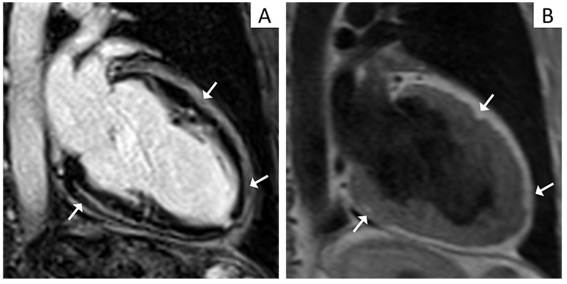
Figure 2.
Cardiac magnetic resonance (CMR) imaging features of a patient affected by left-dominant arrhythmogenic cardiomyopathy. Post contrast CMR images in two-chamber view showing extensive late gadolinium enhancement in the form of stria proceeding from the epicardium towards the endocardium in the inferior and anterior walls (Panel A, arrows). T1 weighted CMR sequences in two-chamber view evidencing fatty infiltration in the same regions as in Panel A (Panel B, arrows).

