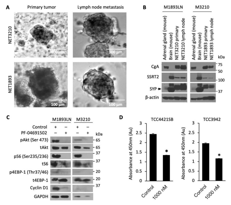
Figure 4.
Patient-derived NET tumor spheroids treatment with PF-04691502. (A) Photographs of tumor spheroids in low-attachment plates. GEP-NET patient tumor sample M3210 was a pT3N2M1a grade 2 tumor ileal neuroendocrine tumor, and tumor sample M1893 was a pT3N2Mx small intestinal neuroendocrine tumor. (B) GEP-NET tumor spheroids were plated in low-attachment plates and 7 d later protein lysates were collected for GEP-NET origin confirmation by Western blot. Mouse brain and mouse adrenal glands were used as positive controls for SYP and CgA, correspondingly. (C) GEP-NET tumor spheroids were seeded in low-attachment plates with equal density and treated with 500 nM PF-04691502 for 24 h. Protein lysates were collected to confirm PI3K/Akt pathway inhibition by pAkt (Ser473), pS6 (Ser235/236) and p4EBP-1 (Thr37/46) by immunoblotting. β-actin was used as a loading control. GAPDH was used as a loading control. (D) GEP-NET tumor spheroids were seeded in a 96-well clear round-bottom ultra-low attachment microplate at 10,000 cells / 1 mL density and treated 24 h later with 1000 nM PF-04691502. CCK-8 reagent was added to each well 48 h later; CCK-8-induced colorimetric changes were measured by absorbance at 450 nM, which is directly proportional to the rate of cell proliferation, after 72 h. * denotes p-value < 0.01.

