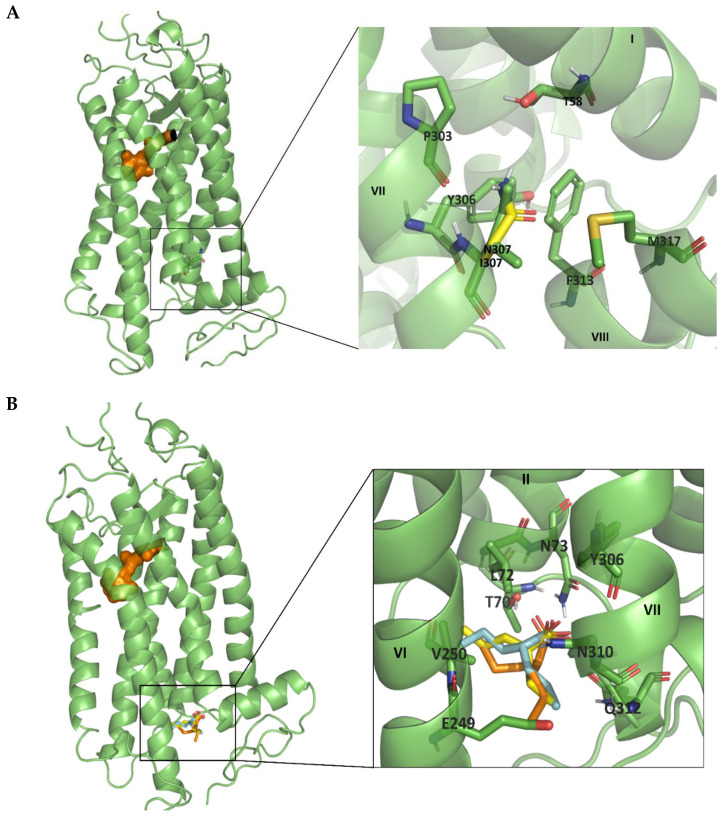
Figure 7.
(A). Structure of Rho receptor, PDB code 1U19, focusing on the position of Ile307. Detailed representation of the Ile307 position and the nearby residues can be observed on the right, where the mutation to Asn is represented in bright yellow. (B). Potential VPA binding site to the intracellular domain of Rho. Positions of the best three poses of VPA docking, with values of the scoring function of −4.5, −4.4, and −4.4 kcal/mol, respectively, are represented in cyan, bright yellow, and orange.

