Figure 1.
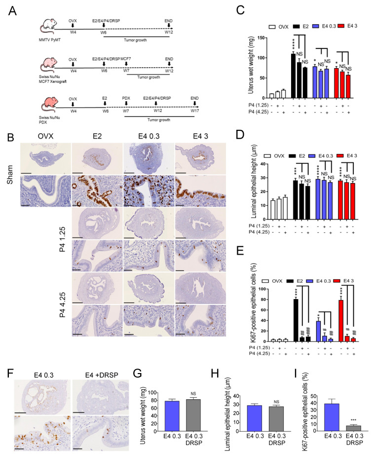
Uterotrophic effect of estetrol (E4), estradiol (E2), progesterone (P4) and drospirenone (DRSP). (A) Treatment protocol schema of the three hormone-dependent breast cancer mouse models: MMTV-PyMT, MCF7 xenograft and patient-derived xenograft (PDX). OVX, ovariectomy; E2/E4/P4/DRSP treatment start pointed by arrows; MCF7, tumor cell injection; PDX, tumor graft; END, mouse sacrifice; W4-W17, 4–17 weeks of age. (B) Representative Ki67 immunostainings on uterus harvested from MMTV-PyMT mice untreated (OVX) or treated with E2, E4 (0.3 or 3 mg/kg/day) combined with or without P4 (1.25 or 4.25 mg/kg/day); scale bar = 500 µm, zoom scale bar = 50 µm. Quantification of (C) uterine wet weight, (D) luminal epithelial height and (E) epithelial cell proliferation (Ki67-positive staining). (F) Representative Ki67 immunostainings on uterus harvested from MMTV-PyMT mice treated with E4 (0.3 mg/kg/day) with or without DRSP (0.06 mg/kg/day); scale bar = 500 µm, zoom scale bar = 50 µm. Quantification of (G) uterine wet weight, (H) luminal epithelial height and (I) epithelial cell proliferation (Ki67-positive staining). Kruskal–Wallis analysis followed by Dunn’s post-tests or Mann–Whitney analysis, n = 6–8 mice/condition. NS: not statistically significant; * or #: p < 0.05; ** or ##: p < 0.01; *** or ###: p < 0.001 and **** or ####: p < 0.0001. * versus OVX, # or NS versus corresponding sham/estrogen-alone treated mice.

