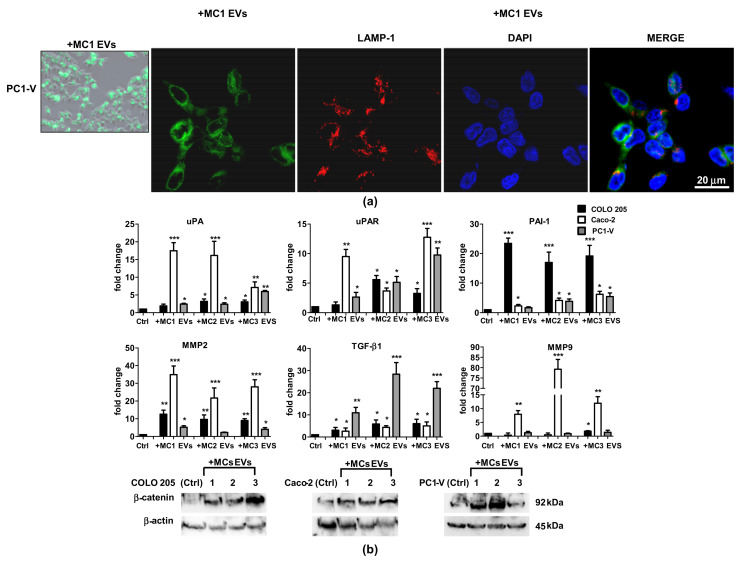
Figure 2.
Uptake of MC EVs causes upregulation of metastatic signaling pathways in tumor cell lines. (a) Left side: Representative images showing the internalization of fluorescent PKH67- MC EVs (green) in the PC1-V cell line (magnification ×400). Right side: Representative images of fluorescence confocal microscopy showing PKH67-MC1 EV uptake and colocalization with LAMP-1 (red) in PC1-V cells (magnification ×1000). (b) Fold change of uPA, uPAR, PAI-1, MMP-2, MMP-9, and TGF-β1 transcript levels in cancer cells after treatment with MC1-3 EVs. Each value reported in the histograms is the mean ± SD of three different experiments performed in triplicate (* p < 0.05, ** p < 0.01, and *** p < 0.001 relative to each untreated control). (c) Representative immunoblots showing β-catenin protein expression in COLO 205, Caco-2, and PC1-V treated with MC1-3 EVs. β-actin is used as loading control.

