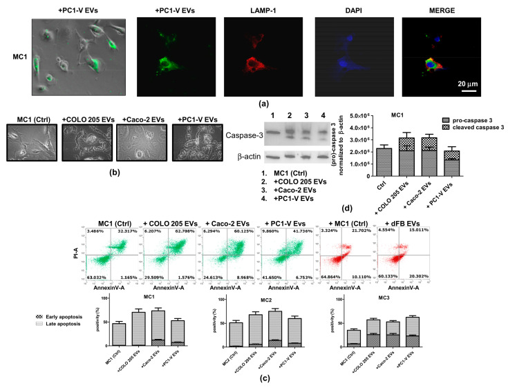
Figure 4.
Uptake of tumor-EVs changes the morphology of mesothelial cells and causes their apoptosis. (a) Representative images of fluorescence and phase-contrast microscopy showing internalization of fluorescent PKH67-PC1-V EVs (green) in the MC1 cell line (magnification ×400) and representative images of fluorescence confocal microscopy showing PKH67-MC EVs uptake and colocalization with LAMP-1 (red) in PC1-V cells (magnification ×1000). (b) Representative images of optical microscopy showing the morphology of MC1 cells before (Ctrl) and after adding tumor EVs. (c) FCM dot plots of AnnexinV/PI positive MC1 cells before and after adding COLO 205 EVs, Caco-2-EVs, and PC1-V EVs and histogram plots depicting the % of early and late apoptosis. Each value reported in the histograms is the mean ± SD of three different experiments. FCM dot plots of AnnexinV/PI positive MC1 cells before and after adding dFB EVs are shown as the non-tumor model. (d) Representative immunoblots and quantification showing the increase in cleaved caspase-3 expression in MC1 cells treated with COLO 205 EVs, Caco-2 EVs, and PC1-V EVs. β-actin is used as a control.

