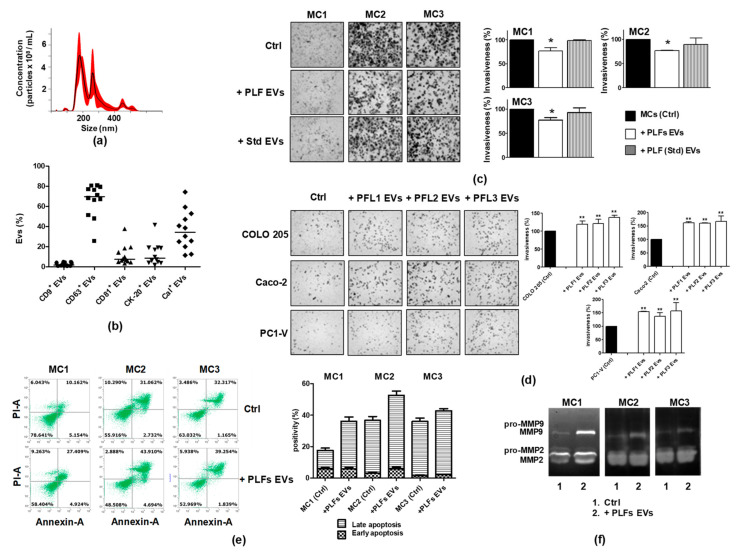
Figure 7.
Biologically active EVs exist in the PLF and are engaged in establishing a pro-metastatic environment. (a) Representative nanoparticle tracking analyses showing the concentration and the size of EVs in the PLF. (b) FCM scatter plot with the median showing coexpression of CK-20 or Cal with the EV markers CD9, CD63, and CD81 in PLF EVs. (c) Representative images showing that a PLF EV pool isolated from a cancer patient reduces the motility of MC1, MC2, and MC3 cell lines, unlike the PLF EV pool from a non-tumor patient (Std) (magnification x100), and quantification of mesothelial cells invasion %. (d) Representative images showing that the PLF EV pool increases the motility of cancer cells and the quantification of tumor cells invasion %. Each value reported in the histograms (c,d) is the mean ± SD of three different experiments performed in triplicate (* p < 0.05 ** p < 0.01 relative to control). (e) FCM dot plots and quantification of AnnexinV/PI positive cells before and after applying a PLF EV pool to MC1-3, showing mainly induction of late apoptosis. (f) Representative gelatin zymography reveals that the addition of a PLF EV pool increases the secretion of active MMP-2/MMP-9 by MC1-3 cells.

