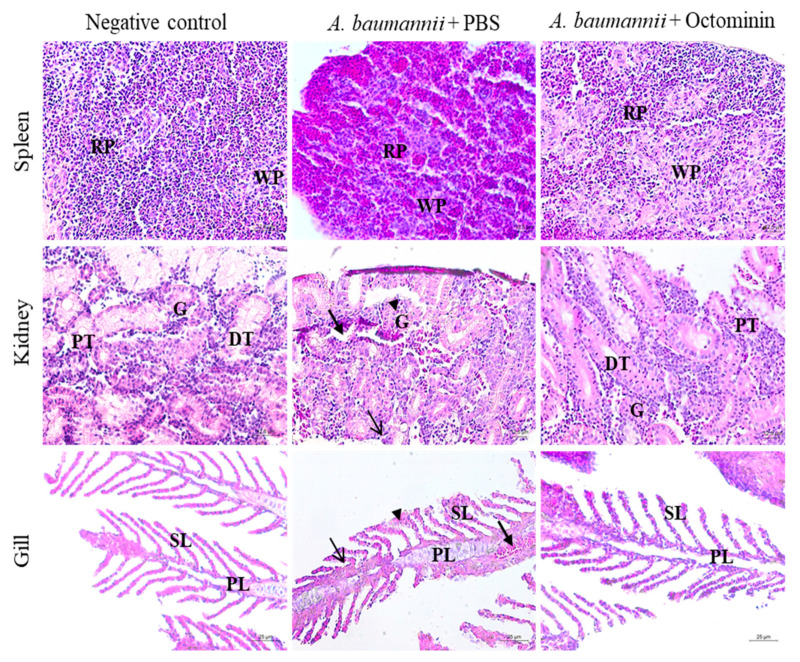
Figure 9.
Histological analysis of zebrafish spleen, kidney, and gill in the negative control, A. baumannii-infected, and PBS-treated, A. baumannii-infected, and Octominin-treated groups. The uninfected groups showed no deviation from the normal structure in all three tissue samples. However, A. baumannii-infected zebrafish spleen had a relatively higher amount of red pulp (RP) than white pulp (WP), while the Octominin-treated sample showed a regular arrangement of the spleen tissue. Kidney samples of A. baumannii-infected and PBS-treated zebrafish had shrunken glomeruli (G), with an increased Bowman space (arrowhead), macrophage infiltration (thin arrow), and cellular necrosis (thick arrow). However, the Octominin-treated group showed fewer deviations, with regular glomeruli, proximal tubule (PT), distal tubule (DT) and a few macrophages. The gill tissue showed erythrocyte infiltration (thick arrow), thickening in primary lamella (PL) (thin arrow), and secondary lamella (SL) clubbing (arrowhead) in A. baumannii-infected and PBS-treated zebrafish, while the Octominin-treated zebrafish showed comparatively fewer deviations. Scale bar represents 12.5 µm in spleen and kidney tissues and 25 µm in gill tissues.

