Figure 2.
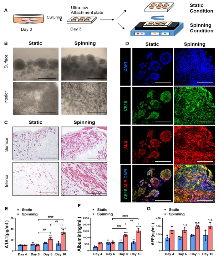
Encapsulated cells in bioprinted hepatic constructs revealed increased proliferative potential and further compacted liver parenchyma under spinning compared to static conditions. (A) Schematic diagram of bioprinted hepatic constructs. Rotatory culture condition generated by orbital shaker was designated as “spinning”, whereas the condition without rotating was referred to as “static”. Rotation was performed at 60 rpm. (B) Morphology of encapsulated cells located at the surface and interior areas of bioprinted hepatic constructs on day 14. Scale bar = 500 μm. (C) Representative H&E staining images showing the localization of encapsulated cells at the surface and interior areas of bioprinted hepatic constructs under static and spinning conditions on day 14. Scale bar = 500 μm. (D) Representative immunofluorescence images of bioprinted hepatic constructs. Sections were stained with cytokeratin 18 (green) and albumin (red) antibodies. Hepatic expression of HepG2 cells within bioprinted hepatic constructs was visualized on day 14 of culturing under static and spinning conditions. Scale bar = 250 μm. (E–G) ELISA for the secretion level of (E) human alpha-1 antitrypsin, (F) human albumin and (G) human alpha-fetoprotein in bioprinted hepatic constructs at indicated time points. The level was calculated every two days from days 4 to 10. Error bars represent the means ± S.D. from three separate experiments. One-way ANOVA followed by Bonferroni’s test was used for the statistical analysis. ** p < 0.01, *** p < 0.001 and **** p < 0.0001 show significant difference between day 4 and another day under the described culture condition. ## p < 0.01, ### p < 0.001 and #### p < 0.0001 indicate significance difference among each day under spinning condition. n.s: no significance.

