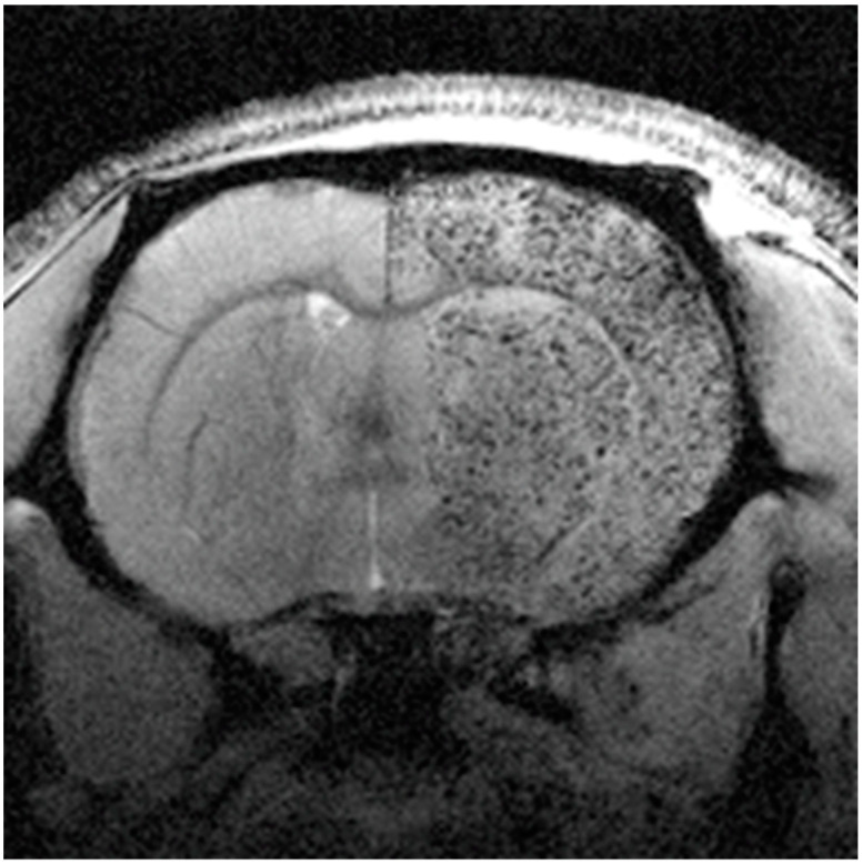
Figure 11.
Magnetic resonance image (MRI) of a rat brain slice, 4 h after intraarterial administration of mesenchymal stem cells (MSCs) labeled with magnetic particles. Labeled cells can be observed as black punctate patterns in the right brain hemisphere. Reproduced with permission from [97], SpringerNature, 2017.

