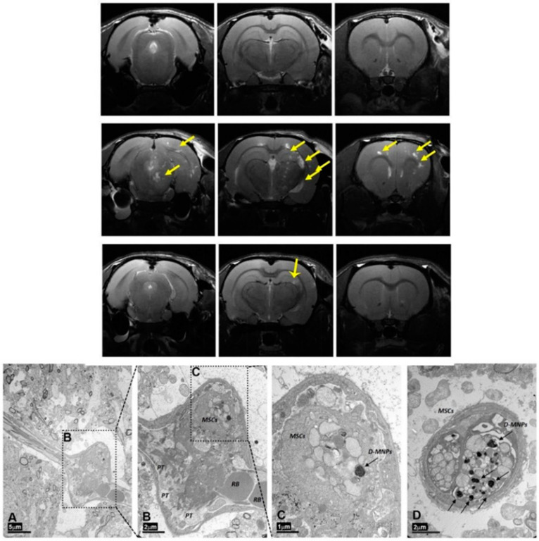
Figure 12.
Upper figure: Multifocal ischemia is observed across the brain after administration of 1 × 10⁶ MSCs (indicated with yellow arrows). Lower figure. Electron transmission micrograph of the rat brain cortex 4 h after intra-arterial (i.a.) delivery of MSCs with magnetic particles, showing the arterial occlusion caused by the cell administration. (A) Dilated brain vessel surrounded by neuropils (scale bar 5 μm). (B) Magnification of vessel dilation in A. Red blood (RB) corpuscles are on the right, and platelets (PTs) and MSCs can be observed inside the vessel (scale bar 2 μm). (C) Magnification of the upper vessel expansion in B (scale bar 1 μm). (D) Longitudinal section of a brain vessel in which two labeled cells can be observed (scale bar 2 μm). Reproduced with permission from [97], SpringerNature, 2017.

