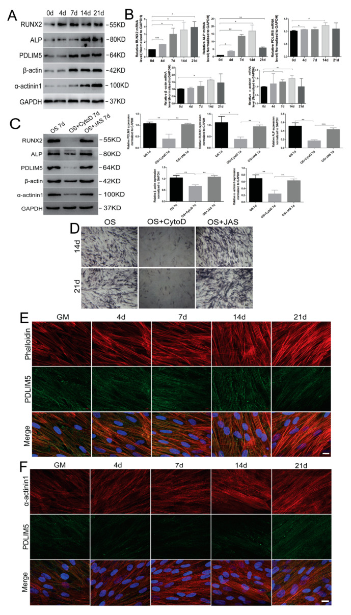
Figure 2.
Microfilaments and related proteins are involved in osteogenic differentiation of HSFs. Detection of β-actin, α-actinin1, PDLIM5, and osteogenesis-associated gene expression by (A) Western blotting and (B) RT-qPCR analyses of HSFs treated with osteogenic differentiation medium (OS) on 0 d, 4 d, 7 d, 14 d, and 21 d. Effects of CytoD and JAS on the expression of HSFS osteogenic-associated protein and PDLIM5 by (C) Western blotting analyses. OS 7d, OS + CytoD, and OS + JAS 7d: HSFs treated with osteogenic differentiation medium, osteogenic differentiation medium containing 0.1 μg/mL of CytoD, and osteogenic differentiation medium containing 10 nM/mL of JAS for 7 days, respectively. Densitometric quantification of the Western blotting bands normalized to GAPDH. * p < 0.05, ** p < 0.01, *** p < 0.001, n = 3. Osteogenic differentiation of HSFs with CytoD or JAS in OS was determined by (D) ALP staining at 14 and 21 days. Scale bar = 100 µm. Immunofluorescence (E) detected the co-localization of PDLIM5 and F-actin, and (F) analysis detected that PDLIM5 co-localized specifically with α-actinin1 on key stress fibers during osteogenic differentiation. Scale bar = 10 µm. Phalloidin: F-actin immunofluorescent dye; CytoD: inhibitors of actin polymerization; JAS: actin polymerization agonist.

