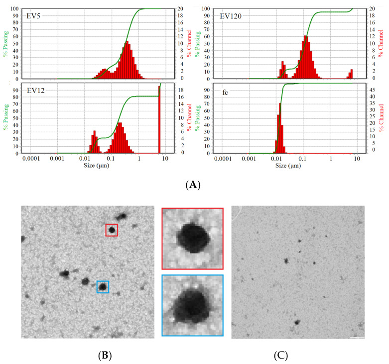
Figure 2.
(A) Particle size distribution by dynamic light scattering of EV5, EV12, EV120 and fc fractions isolated from pools of citrate plasma samples. (B) Transmission electron microscopy of EV120-enriched fraction, two EVs at five-fold enlargement (blue and red box) showing typical EV-like structures of approximately 100 nm diameter and (C) fc fraction.

