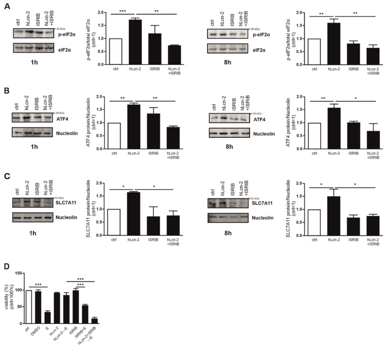
Figure 5.
hLcn-2 delays ferroptosis through induction of the integrated stress response. (A–C) Western analysis of (A) p-eIF2α (n = 3), (B) ATF4 (n = 3), and (C) SLC7A11 protein expression after the co-stimulation of hLcn-2 (5 µg/mL) and the inhibitor of the integrated stress response (ISRIB; 1 µM) for the indicated time points. Either total eIF2α or nucleolin was analyzed as loading control. A representative picture (left panels) from 4 independent experiments is given along with the densitometrical analysis (right panels) (n = 3). (D) Cell viability measured with CellTiter Blue assay, normalized against the unstimulated control. DMSO served as solvent control. Erastin (10 µM) was used to induce ferroptosis for 24 h. CAKI1 cells were co-stimulated with 5 µg/mL hLcn-2 and 1 µM ISRIB for 24 h, washed, and incubated with erastin (10 µM) for additional 24 h (n = 4). Graphs are displayed as means ± SEM with * p < 0.05, ** p < 0.01, *** p < 0.001.

