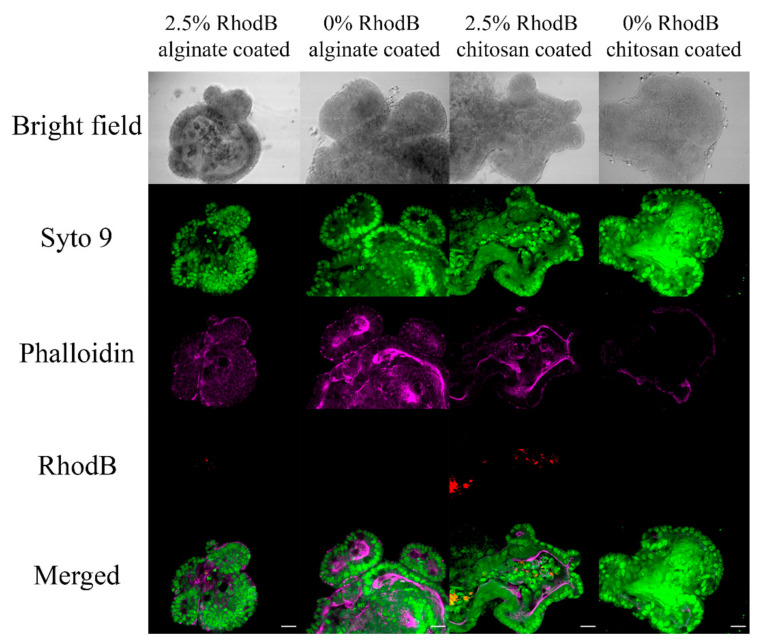
Figure 9.
After adding 2.5% loaded PLGA nanoparticles coated with Alginate and Chitosan (1:4 Nanoparticle: Matrigel), laser microscopy of organoids was added. Green represents SYTO™ 9, magenta represents phalloidin, and red represents Rhodamine B. The magnification of images is 40× with a digital zoom of 1.8, and scale bars represent 20 μm.

