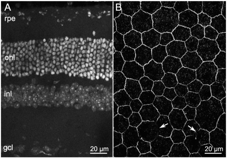
Figure 1.
Retinal and RPE morphologies in wild type (wt) mice aged 1 year. (A) Retinal vertical section, DAPI nuclear staining. (B) Whole-mount RPE, ZO-1 immunostaining. Arrows show short and rare interruptions of the RPE (hexagonal) array. In this and in other Figures: onl—outer nuclear; inl—inner nuclear; and gcl—ganglion cell layer.

