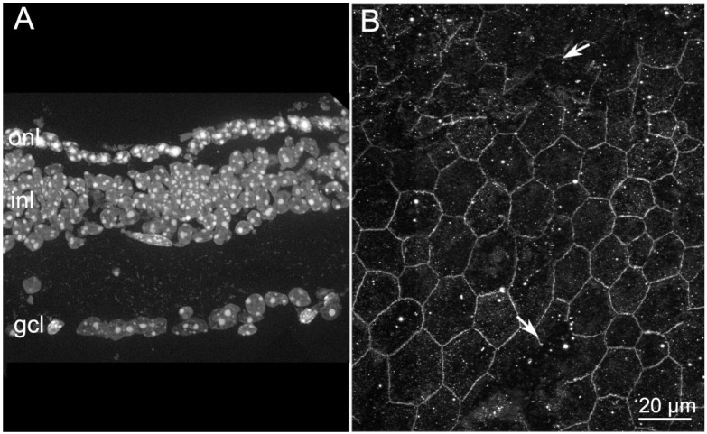
Figure 3.
Retinal and RPE morphologies in rd10 mice aged 45 days. (A) DAPI nuclear staining of retinal vertical sections. Note the persistence of only one row of nuclei in the ONL. (B) ZO-1 staining of RPE from a similar 45 day old mouse in which several wide discontinuities can be appreciated (arrows). Both images are obtained from the central retina/RPE.

