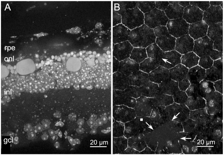
Figure 4.
Retinal and RPE morphologies in light-induced Tvrm4 mice. (A) DAPI nuclear staining of retinal vertical sections from Tvrm4 mouse one week post light-induction. Note the presence of gigantic apoptotic bodies in the ONL, which is composed of few disorganized rows of nuclei compared with the wt. (B) ZO-1 staining of RPE from Tvrm4 mouse one week post light-induction. Arrows show wide discontinuities in ZO-1.

