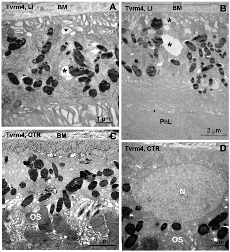
Figure 10.
Vacuolization of RPE cells. Ocular vertical section from Tvrm4 mice, 4 weeks following light-induction (A,B) and in a non-induced littermate (C,D). Transmission electron microscopy shows membranous vacuoles which accumulate at the basal side (abutting Bruch’s membrane, BM) in the proximity of the basal infoldings of RPE cells of the light-induced samples (asterisks in (A,B)); the photoreceptor layer is absent or completely disorganized. In the non-induced control, outer segments (OS) are clearly visible. N—nucleus of RPE cell.

