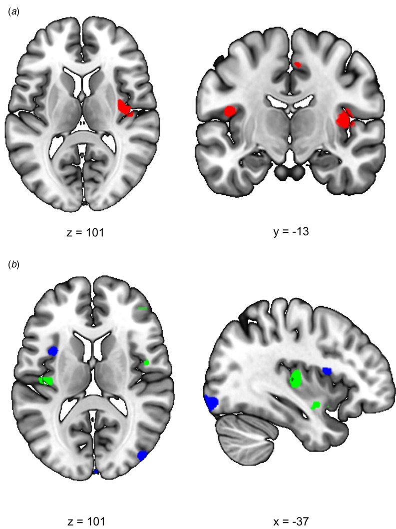
Fig. 2.
Effects of stress on SN functional connectivity in siblings of schizophrenia patients and controls. (a) Group (control/sibling) × stress (stress/no-stress) interaction in SN connectivity during acute stress. Controls and siblings differed in SN connectivity directly after stress (group × stress interaction (psvc < 0.05)) in the right insula. Follow up comparisons revealed that this was driven by a stress-induced increase in healthy controls but not in siblings. (b) Reduced SN connectivity in the aftermath of stress (blue) and a group (control/sibling) × stress (stress/no-stress) interaction (green) in the aftermath of stress. Functional connectivity between the SN and the right anterior insula decreased in the aftermath of stress in both healthy controls and siblings of schizophrenia patients (blue, no-stress > stress psvc = 0.048). Additionally, there was a decrease in functional connectivity between the SN and the posterior insula in the aftermath of stress in siblings (green, group × stress interaction psvc = 0.032 and psvc = 0.057). T-Maps are thresholded at p < 0.001 uncorrected and overlaid onto a normalized anatomical scan for visualization purposes. For a color version, see this figure online. See Table 2 for cluster level inferences.

