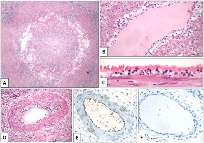
Figure 2.
Spermatic cord arteries show temporal heterogeneity of the lesions, from severe luminal occlusion ((A), original magnification, ×40) to mild endotheliitis with luminal polymorphonuclear elements ((B), ×100) and endothelial tumefaction ((C), ×640). Moderate lesions showed endothelial thickening with mixed inflammatory infiltrates ((D), ×240). Positive immunostaining with SARS-CoV-2 spike antibody was detected in endothelial cells of arteries (((E), ×400) with a negative control in a vein of the same paraffin block ((F), ×400)).

