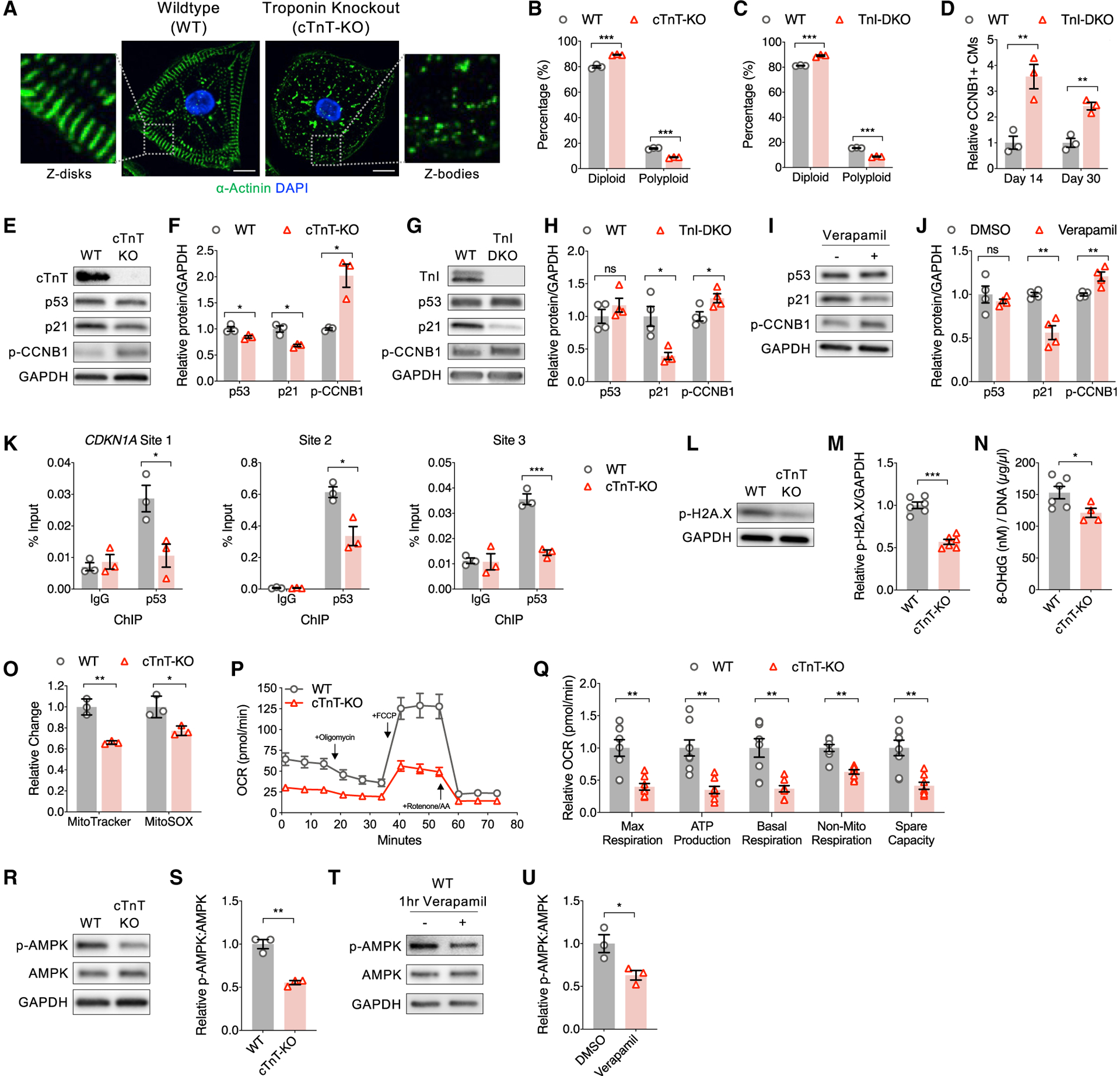
Figure 5. Cellular and molecular consequences of sarcomere assembly.

(A) Representative immunofluorescence images of wild-type (WT) control and cardiac troponin T knockout (cTnT-KO) CMs stained for α actinin (green; sarcomere Z-disk) and DAPI (blue; nuclei). KO of troponin, either cTnT-KO or double KO of skeletal and cardiac troponin I (TnI-DKO), leads to lack of striated sarcomere Z-disks. Scale bar, 10 μm.
(B and C) Flow cytometry of Hoechst-stained CMs demonstrates decreased polyploidy in (B) cTnT-KO and (C) TnI-DKO CMs relative to WT.
(D) Flow cytometry of CCNB1-stained WT and TnI-DKO CMs demonstrates increased proportion of CCNB1+ CMs with TnI-DKO, which reduces with age.
(E and F) Representative immunoblots (E) with (F) quantification of protein lysates from WT and cTnT-KO CMs probed for cTnT, p53, p21, phospho-CCNB1, and GAPDH.
(G and H) Representative immunoblots (G) with (H) quantification of protein lysates from WT and TnI-DKO CMs probed for TnI (cardiac and skeletal), p53, p21, p-CCNB1, and GAPDH.
(I and J) Representative immunoblots (I) with (J) quantification of protein lysates from day 20 WT CMs treated with verapamil from day 9 and probed for p53, p21, p-CCNB1, and GAPDH.
(K) Anti-p53 ChIP-qPCR of WT and cTnT-KO CMs targeting previously reported p53-bound ChIP-seq peaks directly upstream of CDKN1A (Nguyen et al., 2018), which demonstrates decreased p53 binding of CDKN1A in cTnT-KO CMs at three separate genomic sites (see Figure 3D). (L and M) Representative immunoblots probed for phospho-H2AX and GAPDH (L) with (M) quantification demonstrates sarcomere assembly activates a DNA damage response in WT CMs.
(N) Quantification of genomic DNA lysates from WT and cTnT-KO CMs probed via ELISA for 8-OHdG, a marker of oxidative DNA damage.
(O) Flow cytometry quantification of MitoTracker and MitoSOX dyes in WT and cTnT-KO CMs demonstrates reduced mitochondrial content and ROS in cTnT-KO CMs.
(P and Q) Seahorse Mito Stress oxygen consumption rates (OCR) (P) with (Q) quantification shows a decrease in respiration across all parameters in cTnT-KO CMs compared to WT, demonstrating that sarcomere assembly promotes oxidative metabolism.
(R and S) Representative immunoblots (R) with quantification (S) of protein lysates from WT and cTnT-KO CMs probed for p-AMPK, AMPK, and GAPDH.
(T and U) Representative immunoblots (T) with quantification (U) of protein lysates from WT CMs treated with verapamil for 1 h and probed for p-AMPK, AMPK, and GAPDH.
Data are n ≥ 3 and mean ± SEM; significance assessed by t test and defined by p > 0.05 (ns), *p ≤ 0.05, **p ≤ 0.01, and ***p ≤ 0.001. See also Figure S5.
