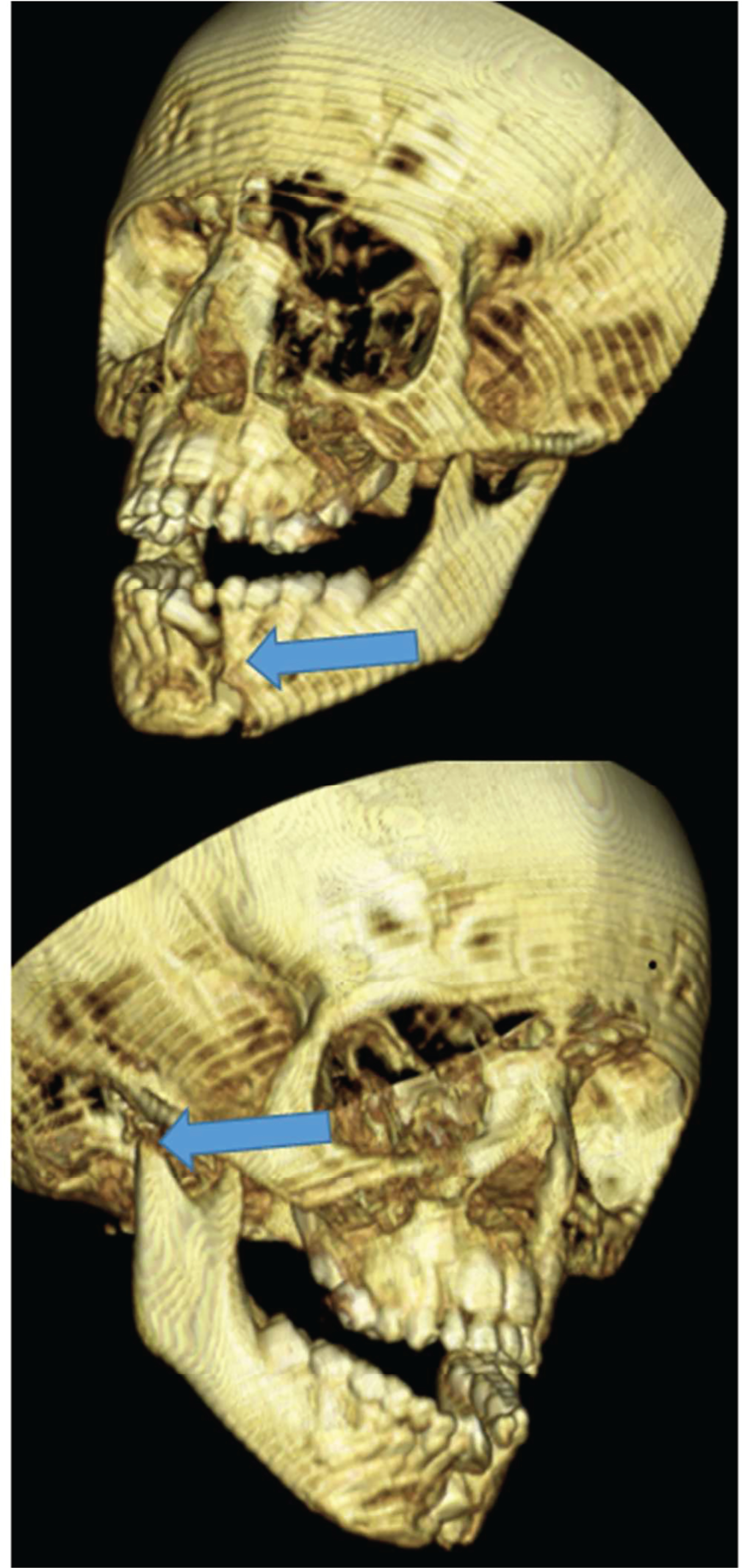
Figure 2.


A: Case 2- 3D CT imaging demonstrating the right mandibular condyle and left parasymphyseal fractures (arrows).
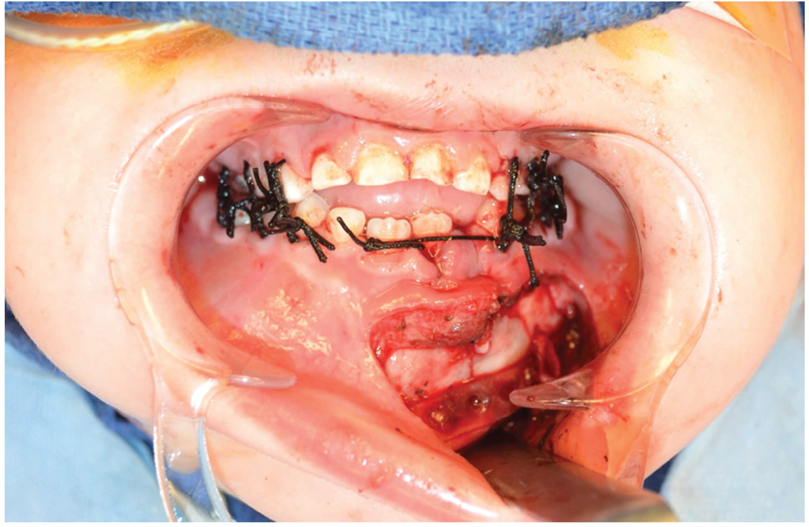
Figure 2B. Intra-operative photo demonstrating intermaxillary fixation with suture ligatures and placement of the absorbable plate over the parasymphyseal fracture.
