Figure 5.


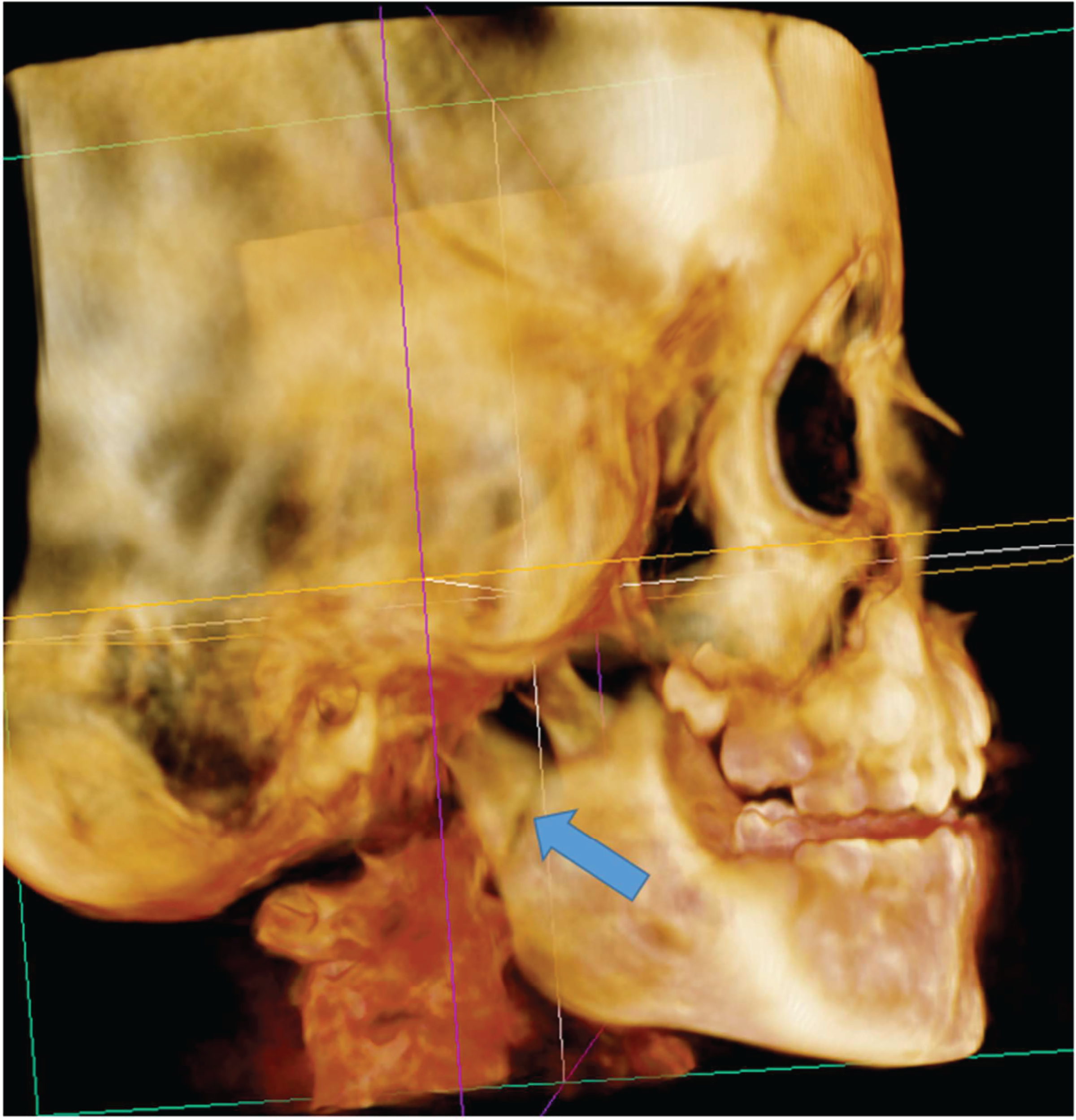
A. Case 5-CT image showing the right subcondylar fracture (arrow).
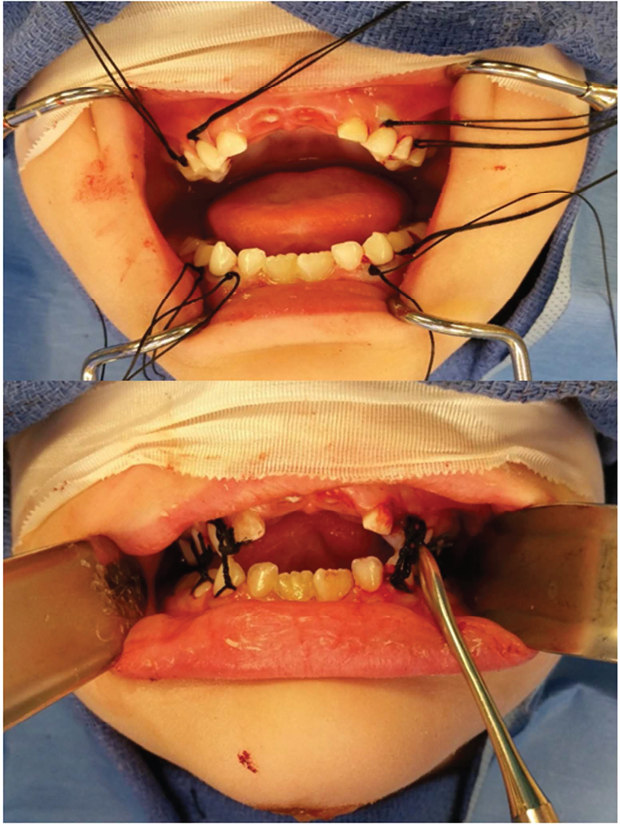
Figure 5B. Demonstration of suture ligatures around the canine and molars (upper) before being tied to each other to provide IMF (lower).


A. Case 5-CT image showing the right subcondylar fracture (arrow).
Figure 5B. Demonstration of suture ligatures around the canine and molars (upper) before being tied to each other to provide IMF (lower).