. 2020 Aug 25;11(35):9675–9684. doi: 10.1039/d0sc03240a

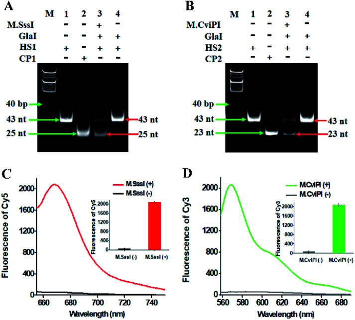
Fig. 1. (A) Nondenaturating PAGE analysis of the cytosine-5 methylation catalyzed by M.SssI and the successive cleavage of HS1 by GlaI. Lane M, the DNA ladder marker; lane 1, the synthesized HS1; lane 2, the synthesized CP1; lane 3, in the presence of M.SssI + GlaI + HS1; lane 4, in the presence of GlaI + HS1. (B) Nondenaturating PAGE analysis of the cytosine-5 methylation catalyzed by M.CviPI and the successive cleavage of HS2 by GlaI. Lane M, the DNA ladder marker; lane 1, the synthesized HS2; lane 2, the synthesized CP2; lane 3, in the presence of M.CviPI + GlaI + HS2; lane 4, in the presence of GlaI + HS2. (C) Fluorescence measurements of RNase HII-mediated recycling liberation of Cy5 molecules from the AuNP nanostructures in the absence (black line) and presence (red line) of M.SssI. Inset shows the fluorescence intensity of Cy5 in the absence (black column) and presence (red column) of M.SssI. (D) Fluorescence measurements of RNase HII-mediated recycling liberation of Cy3 molecules from the AuNP nanostructures in the absence (grey line) and presence (green line) of M.CviPI. Inset shows the fluorescence intensity of Cy3 in the absence (grey column) and presence (green column) of M.CviPI. The 100 U mL−1 M.SssI MTase, 500 U mL−1 M.CviPI MTase, 600 nM HS1, 600 nM HS2, 600 nM CP1 and 600 nM CP2 are used in the experiments.

Fig. 1