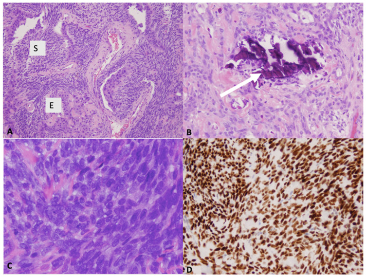
Figure 3.
(A) Microscopic images showing biphasic tumor composed of spindle cell (S) and epithelioid (E) component; H&E ×100 and (B) occasional calcifications (arrow); H&E ×200). (C) Microscopic image showing poorly differentiated morphology (H&E ×400). (D) Immunohistochemistry demonstrating diffuse and strong nuclear staining for the transcriptional corepressor TLE1 (×200).

