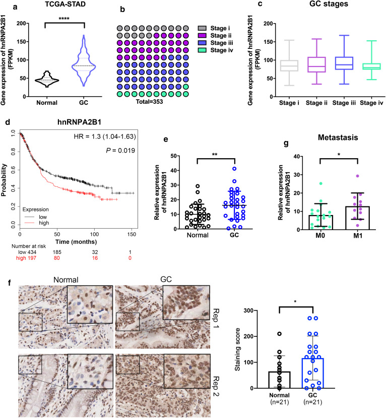
Fig. 2.
hnRNPA2B1 expression is elevated in GC and correlated with adverse prognosis. a The hnRNPA2B1 mRNA expression levels in adjacent non-cancerous tissues and GC tissues from TCGA dataset. b The distribution of neoplasm histologic stages in GC patients of TCGA dataset. c The hnRNPA2B1 mRNA expression in patients with different stages. d Overall survival for GC patients with high (n = 197) and low (n = 434) hnRNPA2B1 expression by Kaplan–Meier curve. e qRT-PCR results of hnRNPA2B1 expression in 29 paired primary GC tissues and adjacent non-cancerous tissues. f Immunohistochemistry staining of hnRNPA2B1 in primary GC tissues and adjacent non-cancerous tissues (n = 21). Significant increased hnRNPA2B1 protein expression in both nucleus and cytoplasm was observed. The score was obtained by multiplying staining intensity (0–3) and the proportion of pituitary cells stained for each intensity to give a value between 0 and 300. g qRT-PCR results of hnRNPA2B1 expression in the above 29 GC tissues with no distant metastases (M0) or with distant metastases (M1). Data are shown as means ± S.D

