Fig. 3.
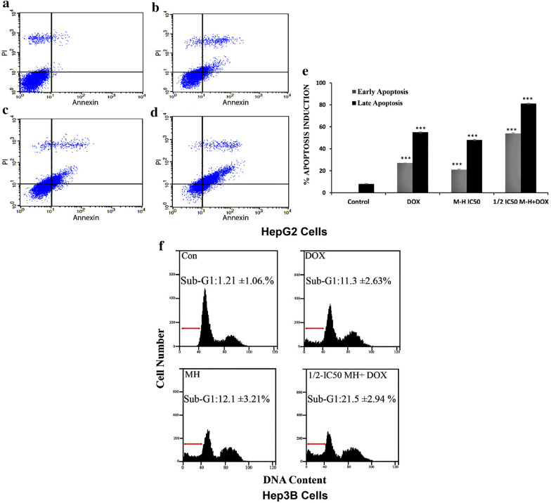
Flow cytometric analysis of early and late apoptosis in treated HepG2 cells with MH, DOX, or combined treatment. HepG2 cells were treated with MH, DOX, or combined treatment for 48 h, stained with FITC-conjugated AV and PI, and subjected to analysis by flow cytometer (a–d). The dot plot represents the untreated HepG2 cells as a control (a) treated cells with 1 μM DOX (b) treated cells with ½ IC50 MH (c) and cells treated with ½ IC50 MH + 1 μM DOX combined treatment (d). The early apoptosis events (AV + /PI−) are shown in, the lower right panels. The late stage of apoptosis (AV + /PI +) is shown in, upper right panels. The bar chart represents the percentage of apoptosis induction in early and late apoptosis in treated HepG2 cells with MH, DOX, or combined treatment. e the results are shown as mean M ± SD of three independent experiments, and Statistical analysis was performed using one-way ANOVA with Tukey’s post-hoc test (*P < 0.05, ** P < 0.01, and ***P < 0.001). f Cell cycle analysis of Manuka honey induced apoptosis in Hep3B cells. Induction of apoptosis by MH or combined treatment with MH and DOX in Hep3B cells. Hep3B cells were exposed to Control, 1 μM DOX, ½ IC50 MH, and ½ IC50 MH + 1 μM DOX combined treatment for 48 h. Cell cycle analysis was performed, quantified for treated-Hep3B cells, and (%) sub-G1 was calculated, using flow cytometry as shown by histograms. The results were obtained from three independent experiments. The figures are representative of one of the experimental results

