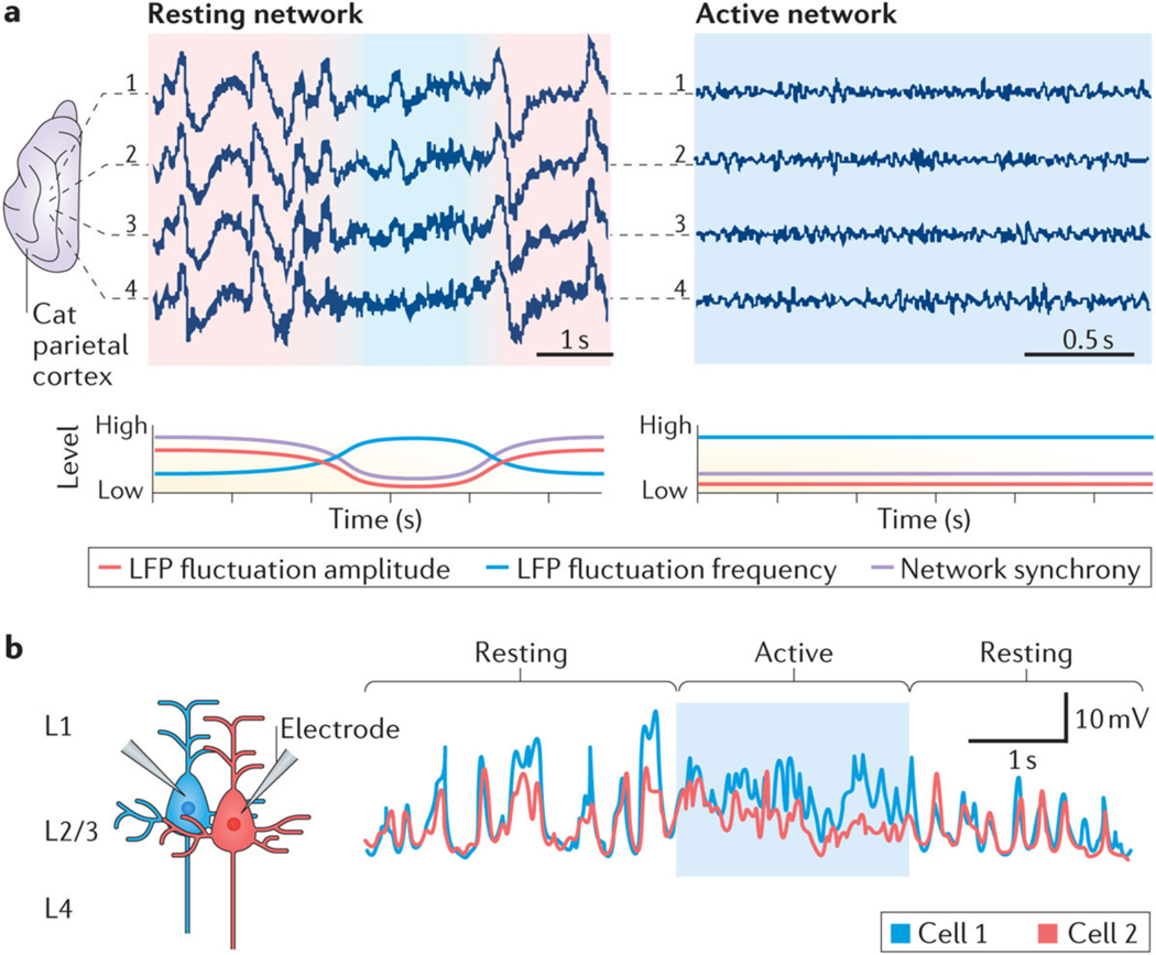
Figure 1 |. Synchrony and functional states of networks.
The degree of correlated neuronal activity (synchrony) reflects the functional state of networks and circuits. a | To illustrate this relationship at the network level, the top panels show local field potentials (LFPs) recorded from four electrodes (1–4) inserted 1 mm apart into the cat parietal cortex (suprasylvian gyrus) during sleep (left) and wakefulness (right). Network activity during resting and non-active states is predominantly characterized by synchronized slow-frequency and high-amplitude fluctuations (red shading). By contrast, network activity during active states is characterized by desynchronized fast-frequency and low-amplitude fluctuations (blue shading). The bottom panels show hypothetical representations of the amplitude (red line) and frequency (blue line) of LFP fluctuations and of the associated network synchrony (pink line). b | Membrane potential recordings of two layer 2/3 (L2/3) pyramidal neurons from mouse parietal cortex (whisker barrel cortex) during resting and active (whisker use) periods illustrate that neurons desynchronize during active periods. Part a is republished with permission of Society for Neuroscience, from Spatiotemporal analysis of local field potentials and unit discharges in cat cerebral cortex during natural wake and sleep states, Destexhe, A., Contreras, D. & Steriade, M., 19, 11, 1999; permission conveyed through Copyright Clearance Center Inc. Part b is from REF.12, Nature Publishing Group.

