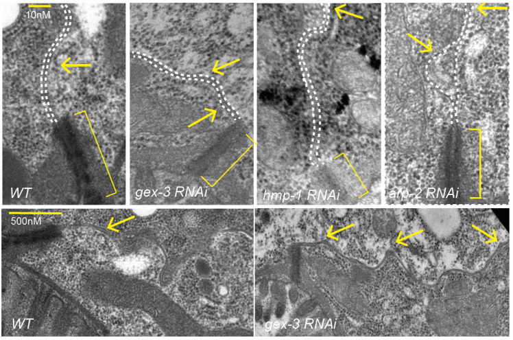
Figure 4.
Electron micrographs (EM) analysis of adult intestines show effect of reducing gex-3 or arp-2 on lateral membranes. Electron micrographs to compare apical and lateral regions of the adult intestine in control animals and animals depleted of components of the WAVE complex (gex-3), Cadherin complex (hmp-1) or Arp2/3 complex (arp-2) via RNAi feeding. Brackets indicate the electron-dense apical junction. Arrows point to the lateral membrane between two intestinal cells, including regions of membrane separation. Dotted white lines follow the lateral membranes between two intestinal cells. * = p < 0.05, ** = p < 0.005, *** = p < 0.0005, ns = not significant.

