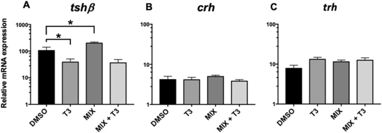
Figure 3.
Changes in relative gene expression by qPCR of (A) thyroid-stimulating hormone-β (tshβ), (B) corticotrophin-releasing hormone (crh), and (C) thyroid-releasing hormone (trh), in tadpole whole brain tissue following exposure to DMSO control, 10 µg/l mixture (MIX), with or without 1.25 nM T3 for 6 days. Data presented include mean and SEM with N = 6. Statistical significance is denoted by * where p < .05 using 1-way ANOVA and Tukey’s post hoc test.

