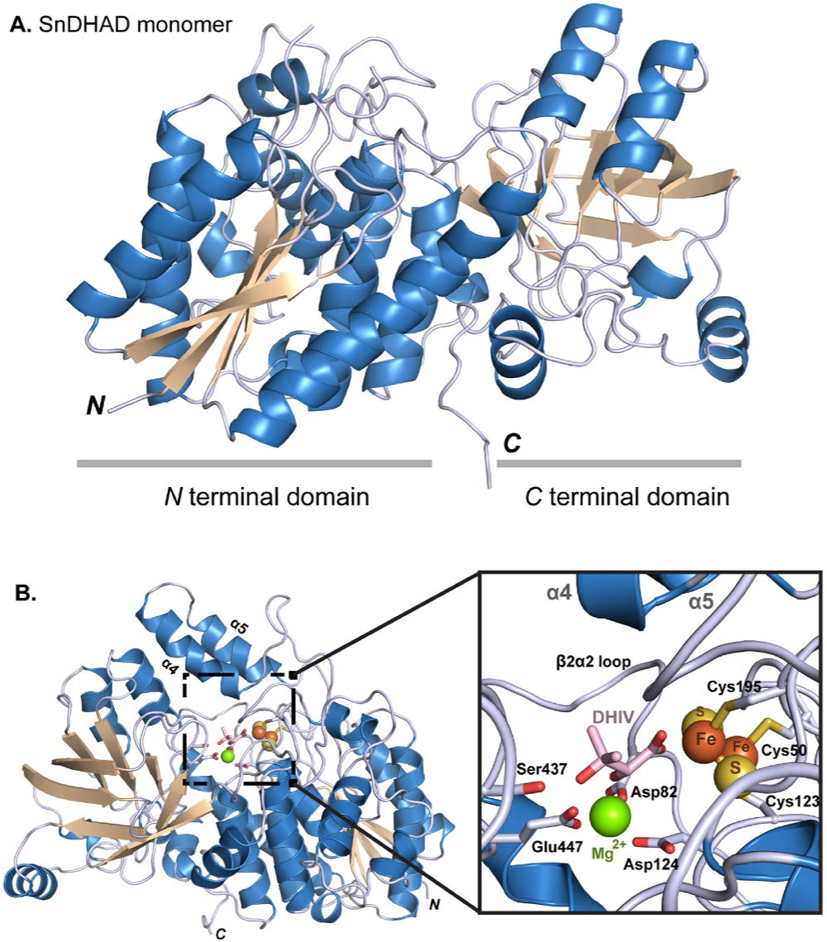
Figure 4.

The crystal structure of SnDHAD. A: Ribbon diagram of the monomer with the two subdomains highlights. B: Ribbon representation of the SnDHAD monomer with modeled active site and substrate DHIV. Expanded view of the active site of SnDHAD showing modeled DHIV coordinated to the [2Fe-2S] cluster and Mg2+.
