
Figure 3.
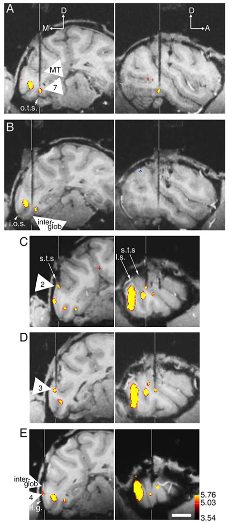
Magnetic resonance images showing the location of five (A-E) tungsten micro-electrode recordings, targeting color-preferring (glob) and non-color preferring (inter-glob) regions of alert macaque brain. Electrodes are black, highlighted by vertical white extension lines. Functional activity (response to equiluminant color > response to achromatic) is superimposed. The brain has been computationally sliced in the plane of the electrode: pseudo-frontal sections (left); pseudo-sagittal sections (right). Numbers relate to the globs identified in Figure 1; approximate A-P coordinates given in Figure 1. Supplementary Figure 4 shows electrodes targeting globs and inter-globs in a second animal. l.g., lunate gyrus; M, medial; D, dorsal; A, anterior; other conventions as for Figure 1. Scale bar is 1 cm.